The work that won the Nobel Prize in chemistry—in terms everyone can understand
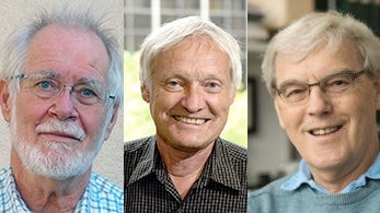
Biology is in the midst of a revolution, and the 2017 Nobel Prize in chemistry has been awarded to the researchers who are at the forefront of innovation in the field. According to the prize citation, Jacques Dubochet (University of Lausanne), Joachim Frank (Columbia University), and Richard Henderson (MRC Laboratory of Molecular Biology in Cambridge, UK) deserve the award for developing “cryo-electron microscopy for the high-resolution structure determination of biomolecules in solution.”


Biology is in the midst of a revolution, and the 2017 Nobel Prize in chemistry has been awarded to the researchers who are at the forefront of innovation in the field. According to the prize citation, Jacques Dubochet (University of Lausanne), Joachim Frank (Columbia University), and Richard Henderson (MRC Laboratory of Molecular Biology in Cambridge, UK) deserve the award for developing “cryo-electron microscopy for the high-resolution structure determination of biomolecules in solution.”
What does it mean?
There are still many things in the natural world that scientists remain utterly clueless about. One fundamental mystery is how complex biological molecules go about creating life in its astonishing diversity. Ask a biologist why they haven’t cracked it yet, and you will likely hear: “structure is function.”
The trouble is that it’s really hard to get a good look at the structure of biomolecules. Such is the challenge that scientists developing tools to improve the resolution and detail of images of crucial molecules often end up winning big prizes. In addition to this year, the chemistry Nobel in 1982, 1985, 1987, and 2002 were given to scientists for developing methods to better determine the structure of molecules. (Separately, the chemistry Nobel in 1958, 1962, 1964, 1972, 1988, and 2009 were won for using molecular structures to determine functions of biomolecules.)

The difficulty overcome by this year’s winners has annoyed scientists for decades. Life began in water, and continues to function in it (remember that 60% of our body weight is water). But water is problematic for structural biologists, who need to stabilize molecules at really low temperatures; frozen water often distorts their view.
Dubochet, Frank, and Henderson developed a method by which water could be frozen really rapidly. The process ensures that water molecules don’t form ice crystals, but instead they just get frozen in place like glass. This vitrification process ensures that biological molecules in the water don’t get distorted.
Their method involves a bow-and-arrow like system that shoots a drop of water containing biological molecules into ethane that has been cooled to -196°C (-320°F). At such temperatures, without ice crystals, biological molecules remain stable enough that you could take an image of them that is no longer blurred.
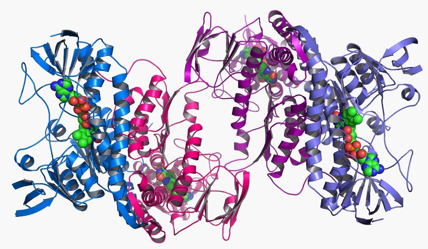
To take those images, Henderson and his colleagues used electron microscopy (EM). This is achieved by shooting beams of electrons at the structure and recording how they are deflected by the underlying molecules.
But standard techniques using EM didn’t produce the kinds of resolution Henderson and his colleagues needed. So they worked with microscope manufacturers to make the process better. By 2012, they had managed to develop the “cryo-EM,” where “cryo” stands for studying biological molecules in a frozen state.
These cryo-EM machines can be as tall as 3 meters (9 feet) and cost more than $7.5 million each. The machine takes thousands of two-dimensional images and stitches them to create a three-dimensional image of molecules. Using the cryo-EM, scientists have been able to image proteins that govern the biological clock and even the Zika virus. Better still, some scientists have been able to create “molecular movies” to study rotor-shaped proteins.
Why does it matter?
With every improved picture of microscopic biological structures, scientists are able to understand better how proteins do their magic. Learning how they work opens all sorts of potential new applications for scientists, most notably identifying new cures for diseases.
There are limitations, of course. Cryo-EM can only be used for large molecules that can withstand being bombarded with electrons. And the technique, though developed over decades, has only been used widely since 2012. It’s also expensive, costing as much as $6,000 per day for running the facility.
Still, a Nobel Prize for cryo-EM is a recognition of what’s in store for structural biologists who use the tool. In a recent example, researchers in the University of California, San Francisco were able to push cryo-EM to image a relatively small protein. “I literally lost an entire night’s sleep when I saw that,” as one researcher put it to Nature.