These photos reveal the hidden worlds around us

“Tulip bud cross section”
Image: Andrei Savitsky
By
We may earn a commission from links on this page.

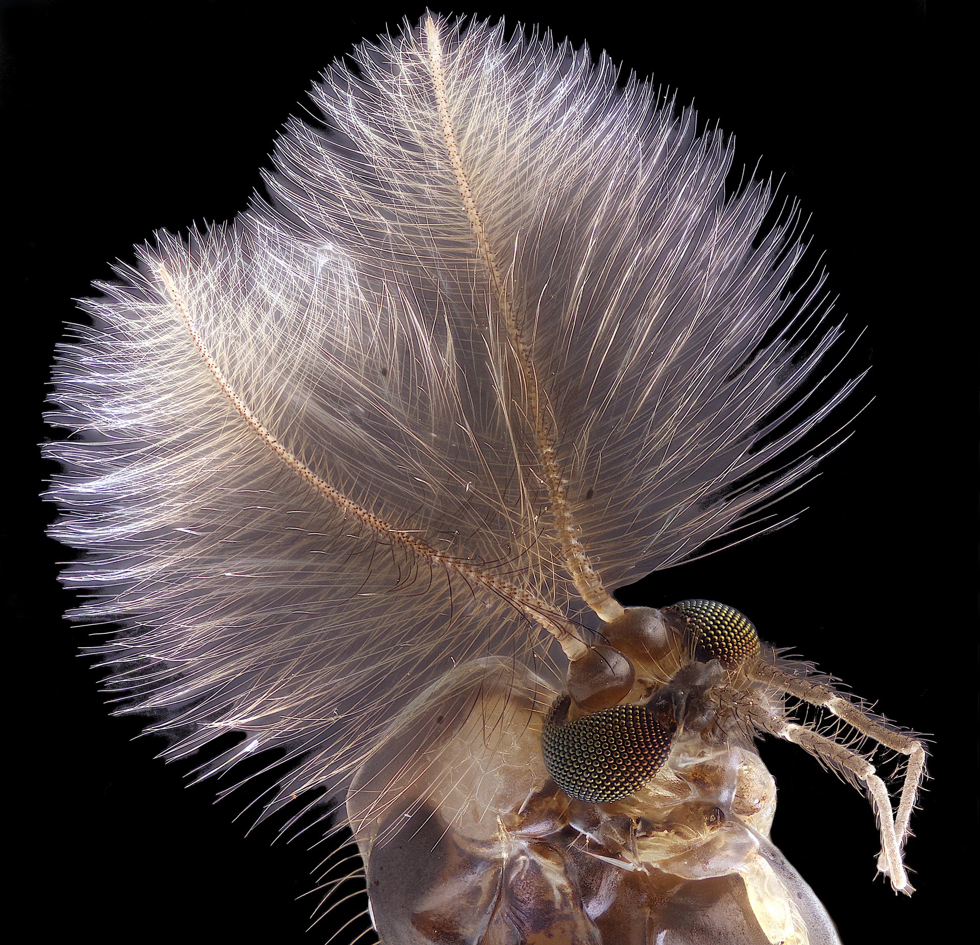
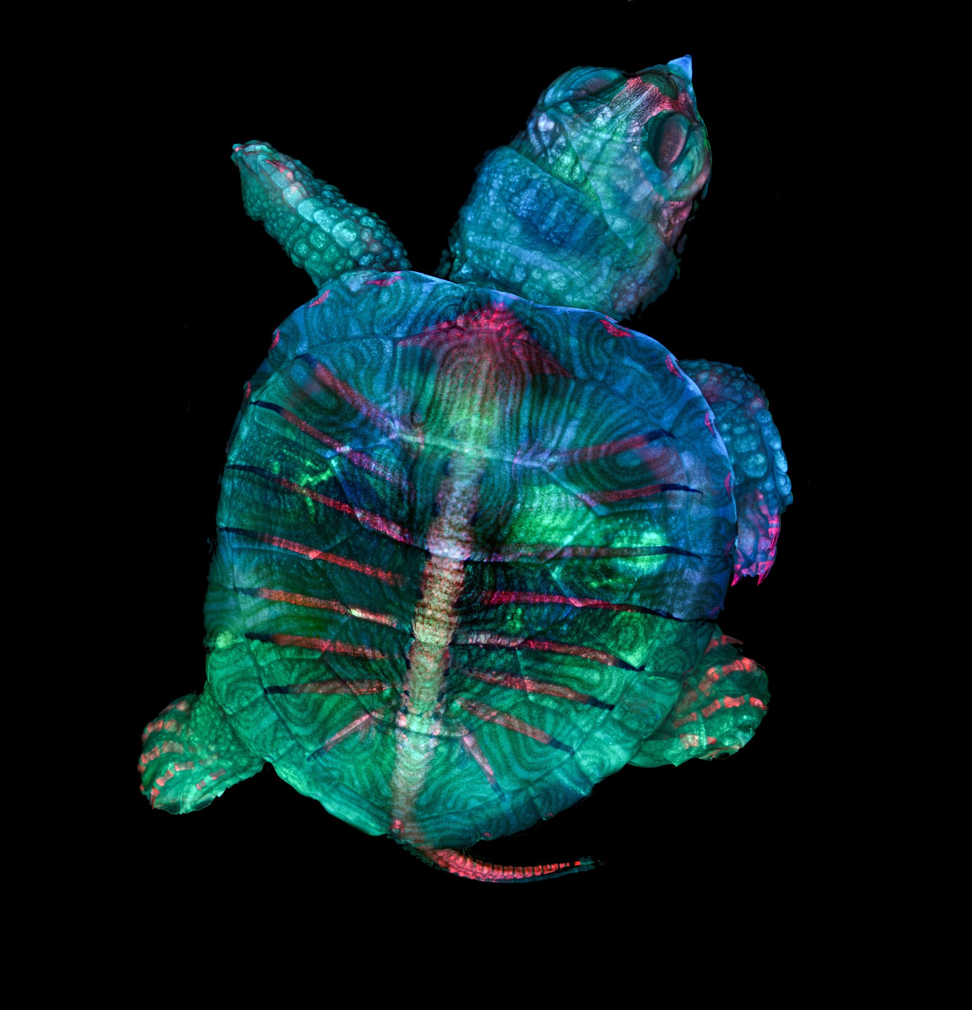
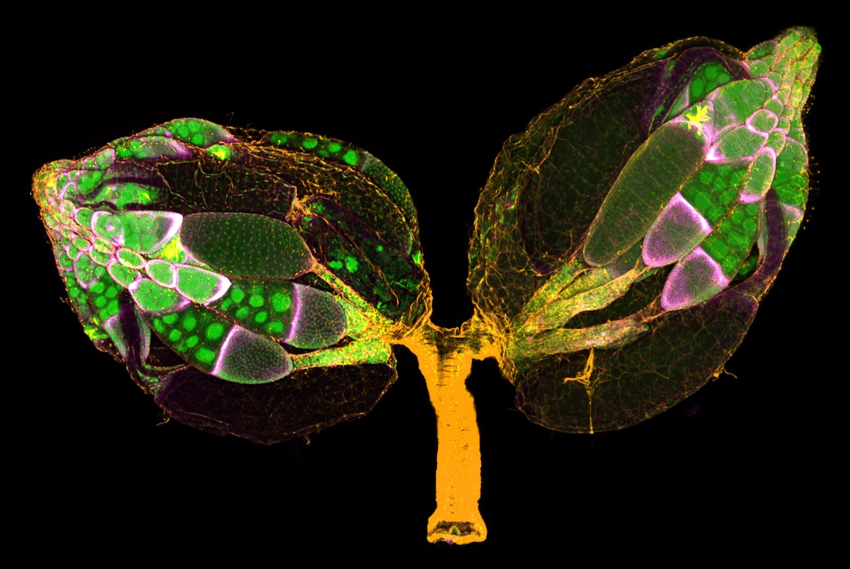
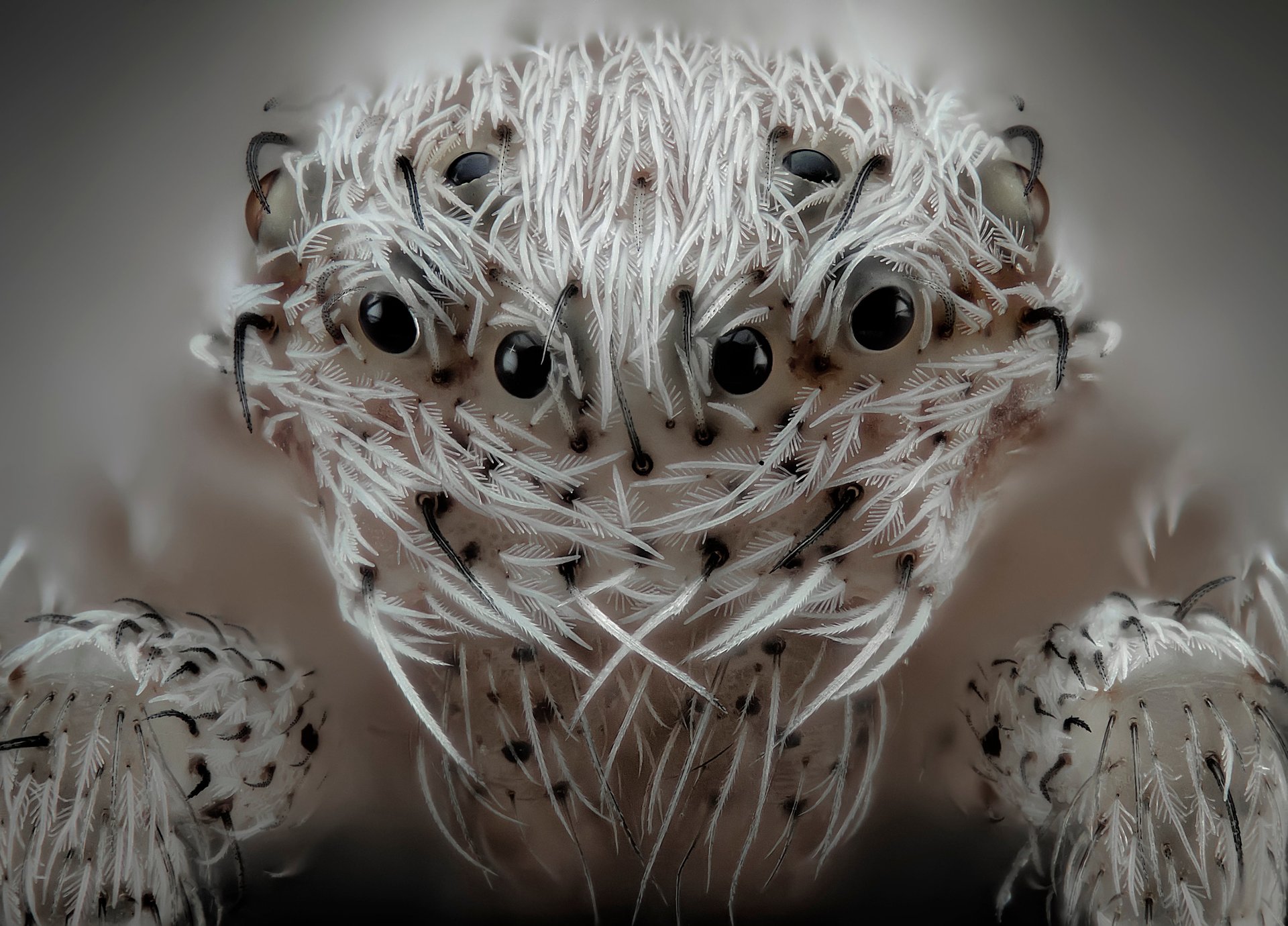
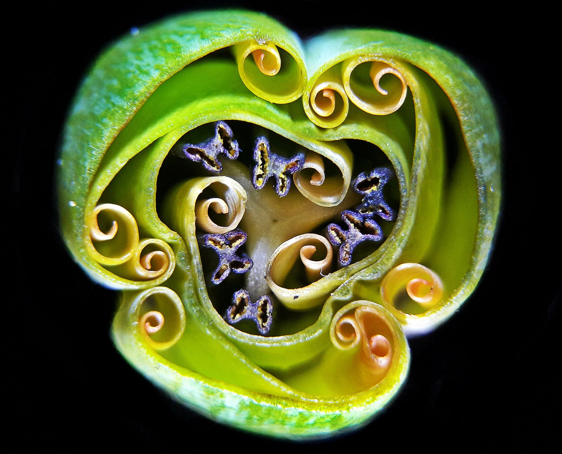
Nikon announced the winners of its Small World microphotography competition today (Oct. 21), awarding amateur and professional photographers (many of whom are also practicing scientists) for pictures that capture the hidden worlds lying at the microscopic level.

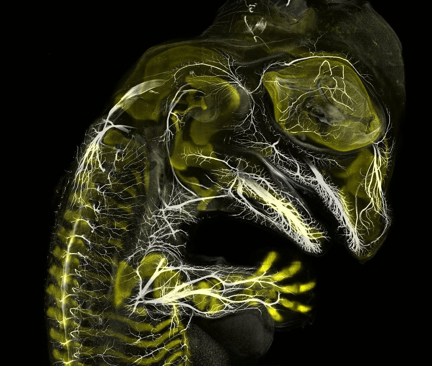
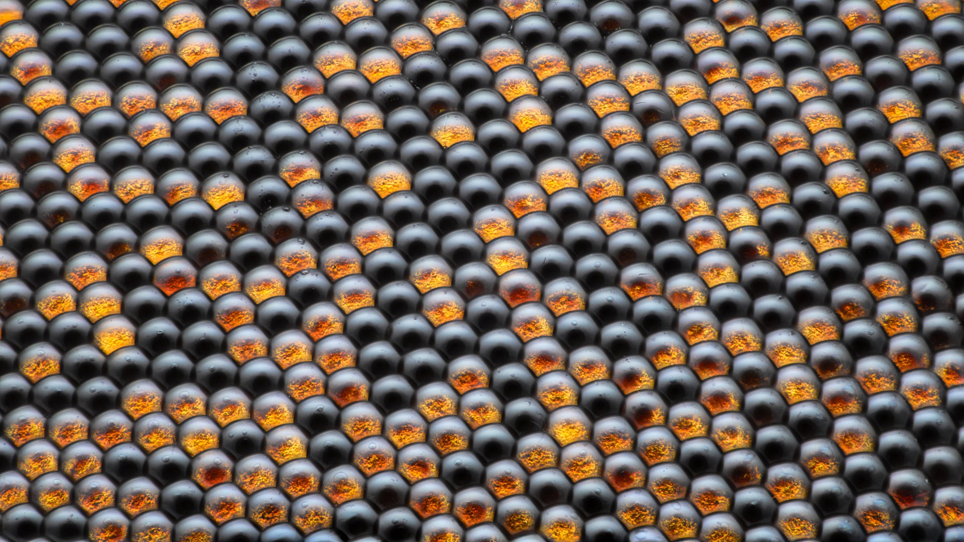
While some images showcase the tiny details not noticeable to most, like the intricate interwoven shapes of a tulip, others delve into the hidden worlds that are impossible to see with the naked eye, like an alligator embryo or the pattern of spheres that make up a housefly’s eye.

Take a look at some of the stunning examples below.

Alligator embryo
Alligator embryo.
Image: Daniel Smith Paredes & Dr. Bhart-Anjan S. Bhullar
Male mosquito
Male mosquito.
Image: Jan Rosenboom
Fluorescent turtle embryo
Fluorescent turtle embryo.
Image: Teresa Zgoda & Teresa Kugler
Tulip bud cross section
“Tulip bud cross section.”
Image: Andrei Savitsky
Housefly compound eye pattern
Housefly compound eye pattern.
Image: Dr. Razvan Cornel Constantin
A pair of ovaries from an adult Drosophila female stained for F-actin (yellow) and nuclei (green); follicle cells are marked by GFP (magenta)
A pair of ovaries from an adult Drosophila female.
Image: Dr. Yujun Chen & Dr. Jocelyn McDonald
Pregnant Daphnia magna (small planktonic crustacean)
Pregnant Daphnia magna (small planktonic crustacean).
Image: Marek Miś
Small white hair spider.
Image: Javier Rupérez