Prize-winning microscopic photography captures the marvels of what we can’t see

Eye of a honey bee (Apis mellifera) covered in dandelion pollen, magnified 120x.
Image: Ralph Claus Grimm/Nikon Small World
By
We may earn a commission from links on this page.

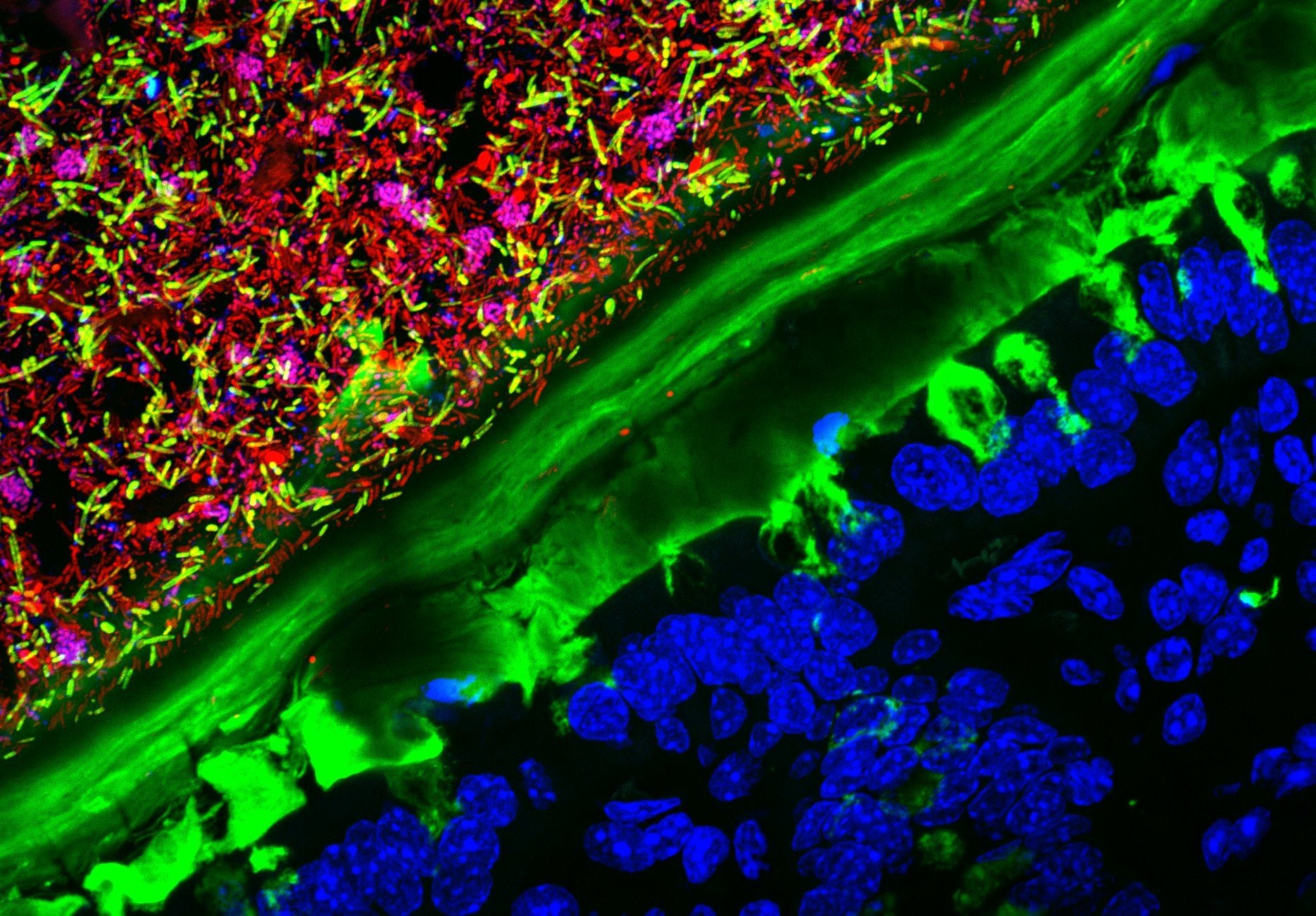
Scientists aren’t the only ones who can experience the beauty of a mouse colon at 63x magnification—and it is beautiful. At high magnification, even a worm or a moth’s antenna can be stunning, and photomicrography makes these wonders of the atomic world visible to everyone.

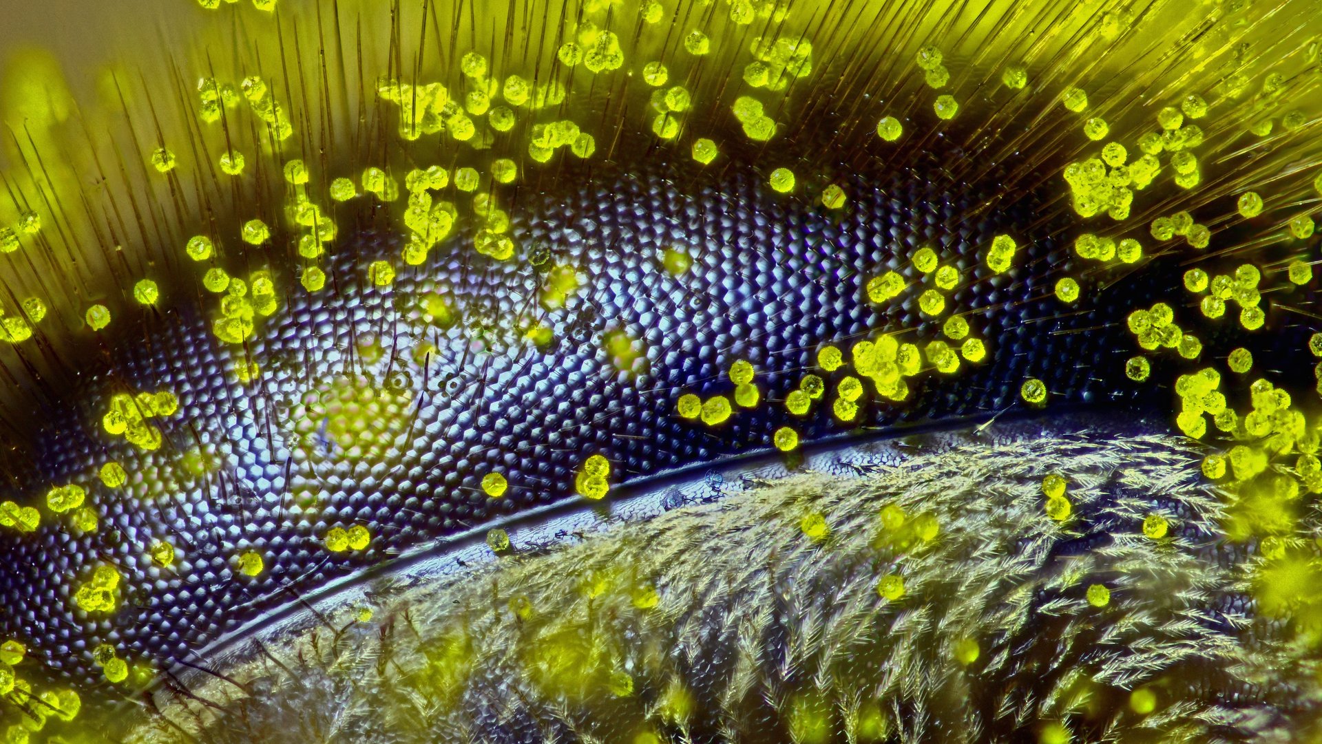
Photomicrography is the art of taking photos through the lens of a microscope. Every year, camera maker Nikon chooses the best photomicrographic images in its “Small World” competition; this year’s winner (above) is a picture of a honey bee’s eye covered in dandelion pollen, taken by Australian high school teacher and beekeeper Ralph Grimm.

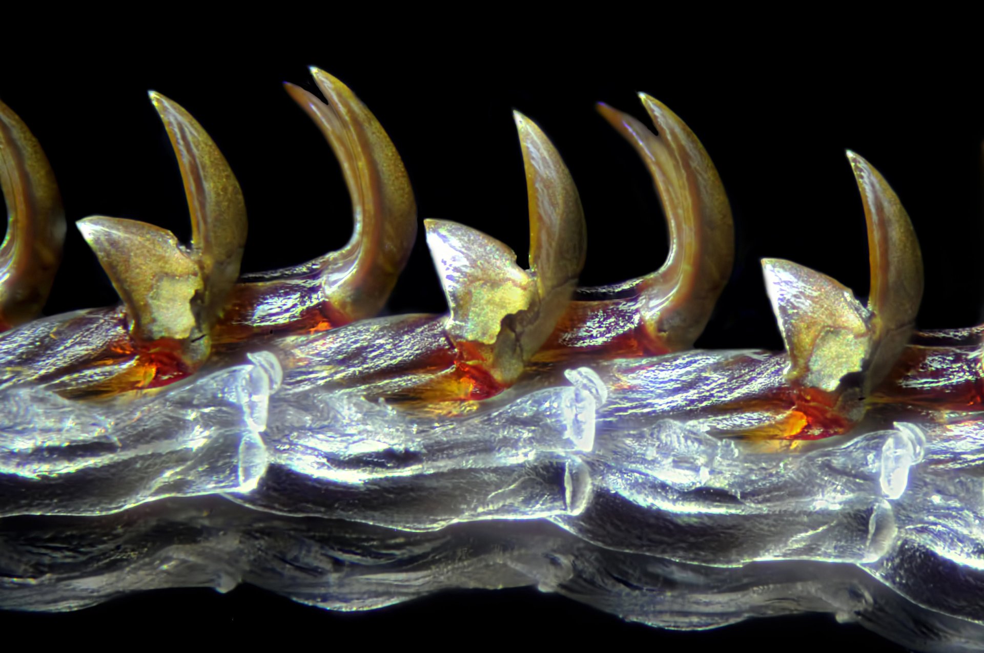
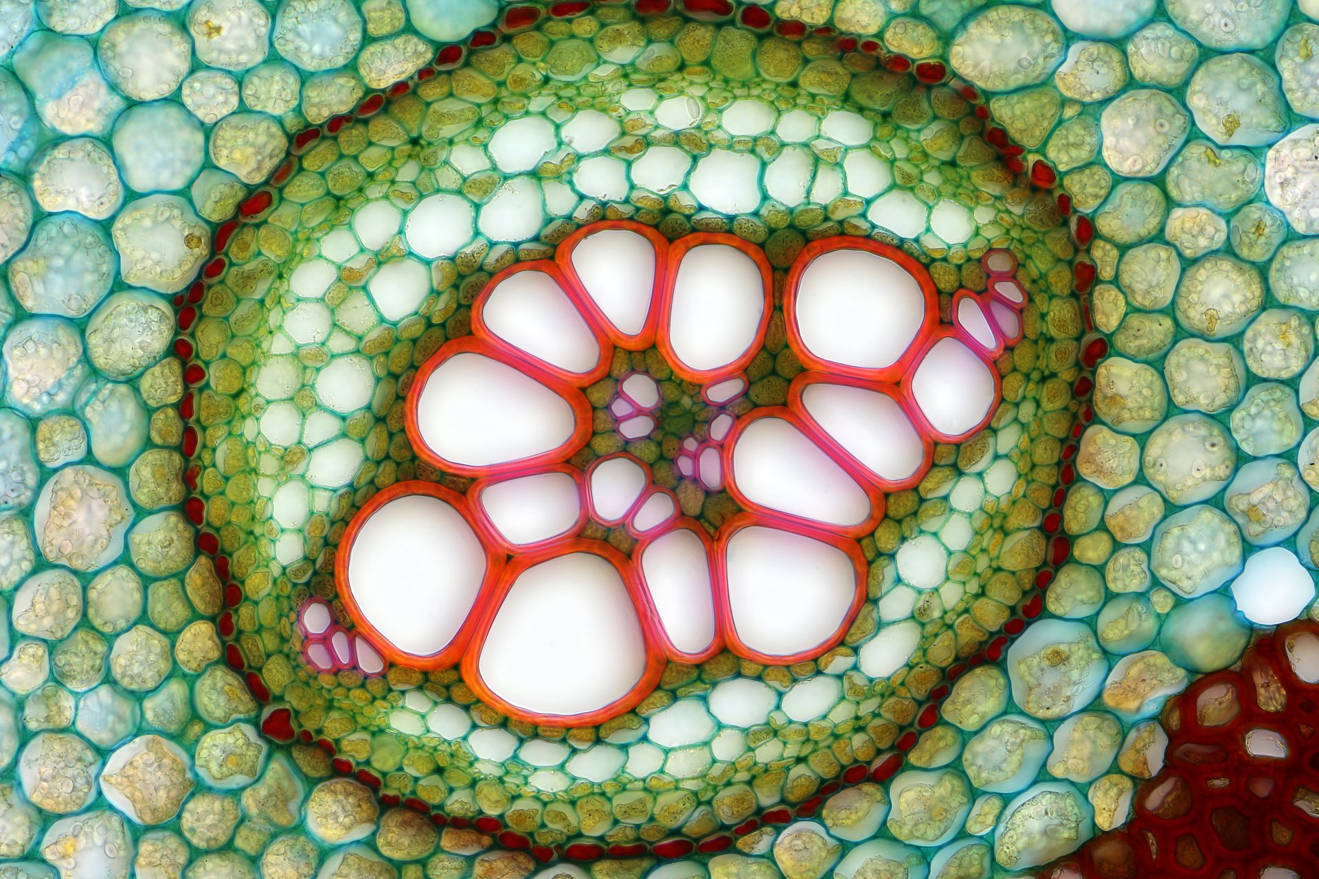
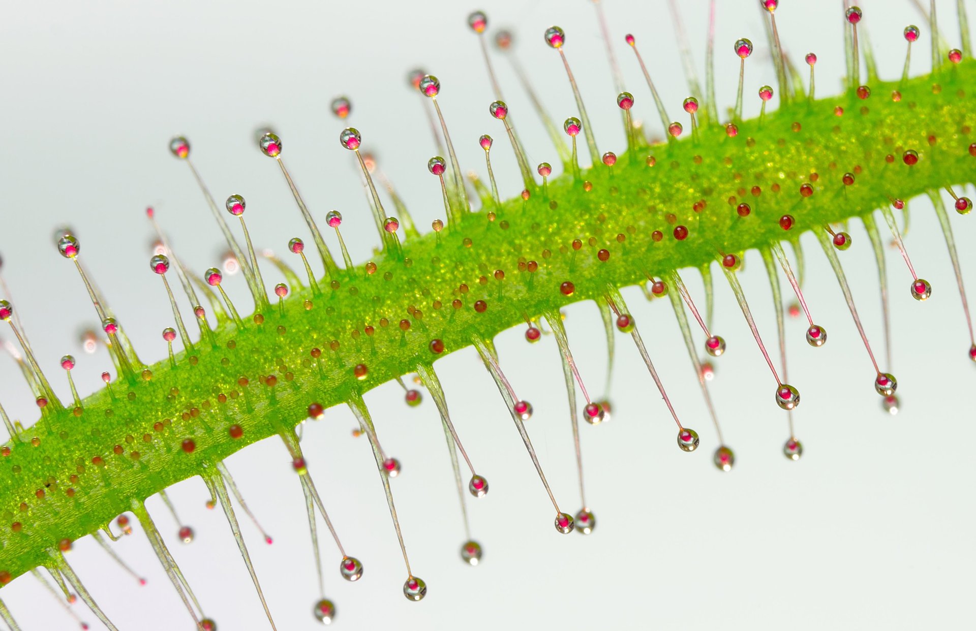
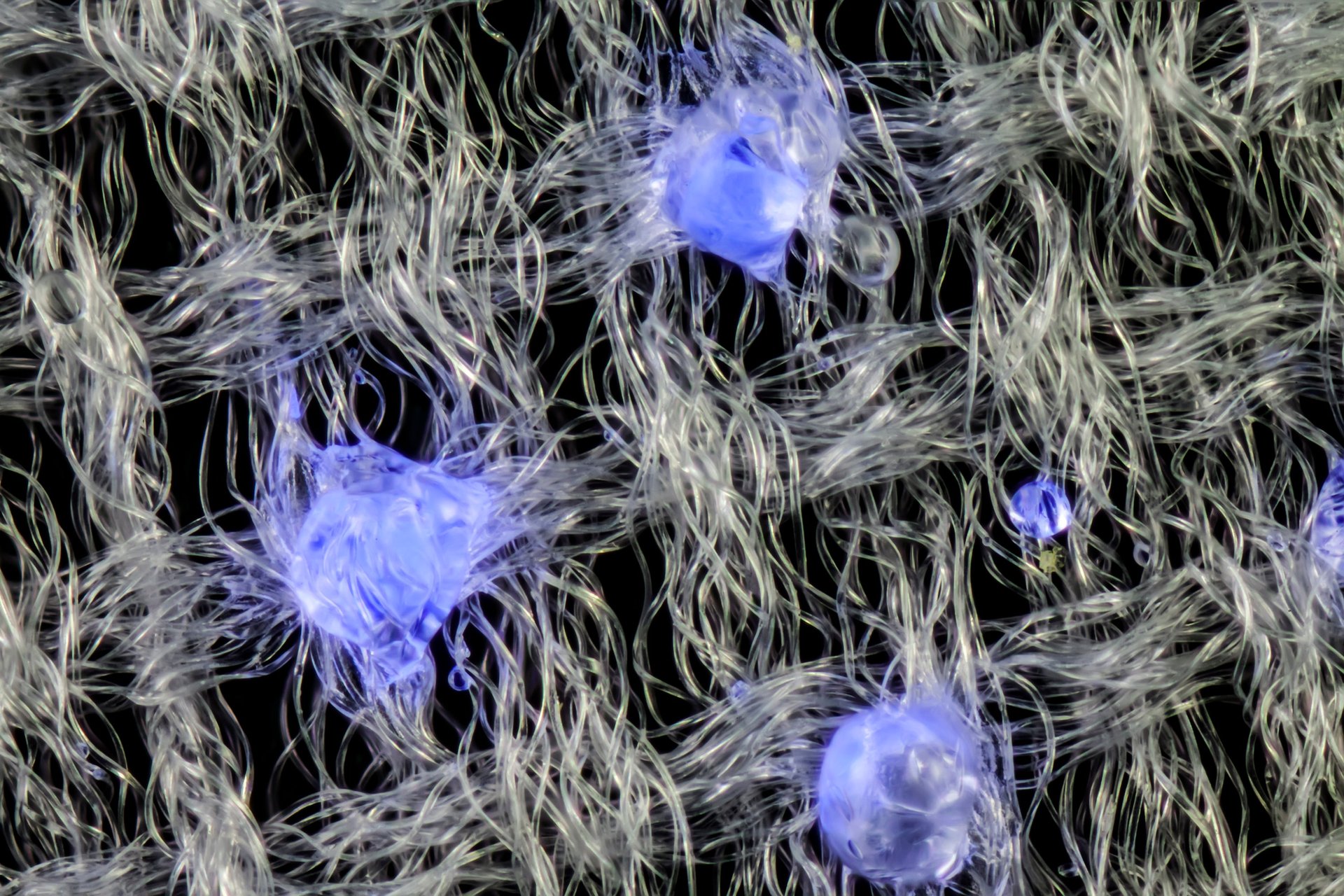
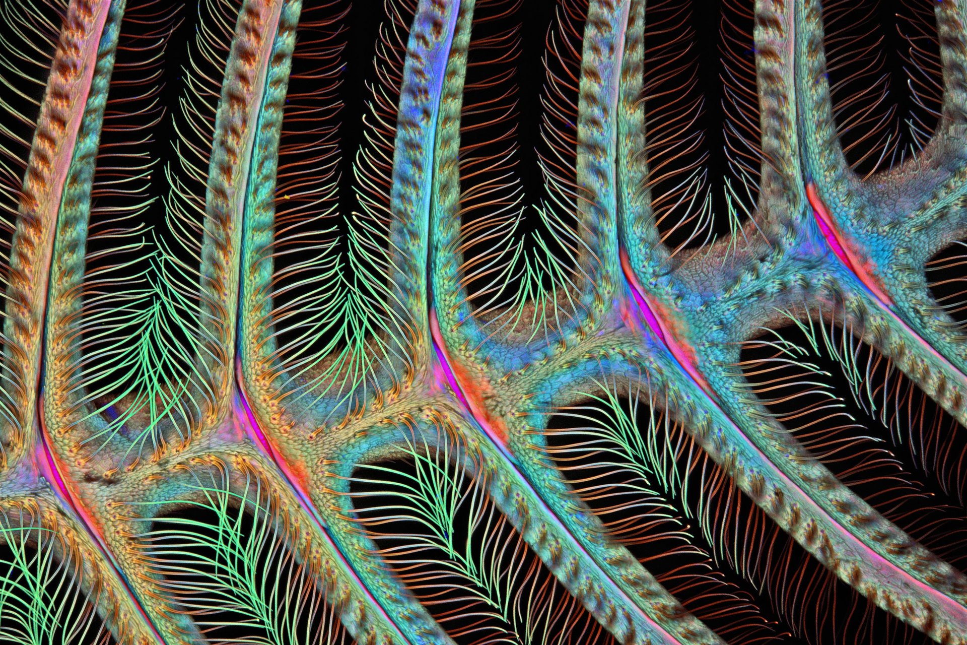
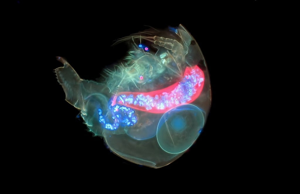
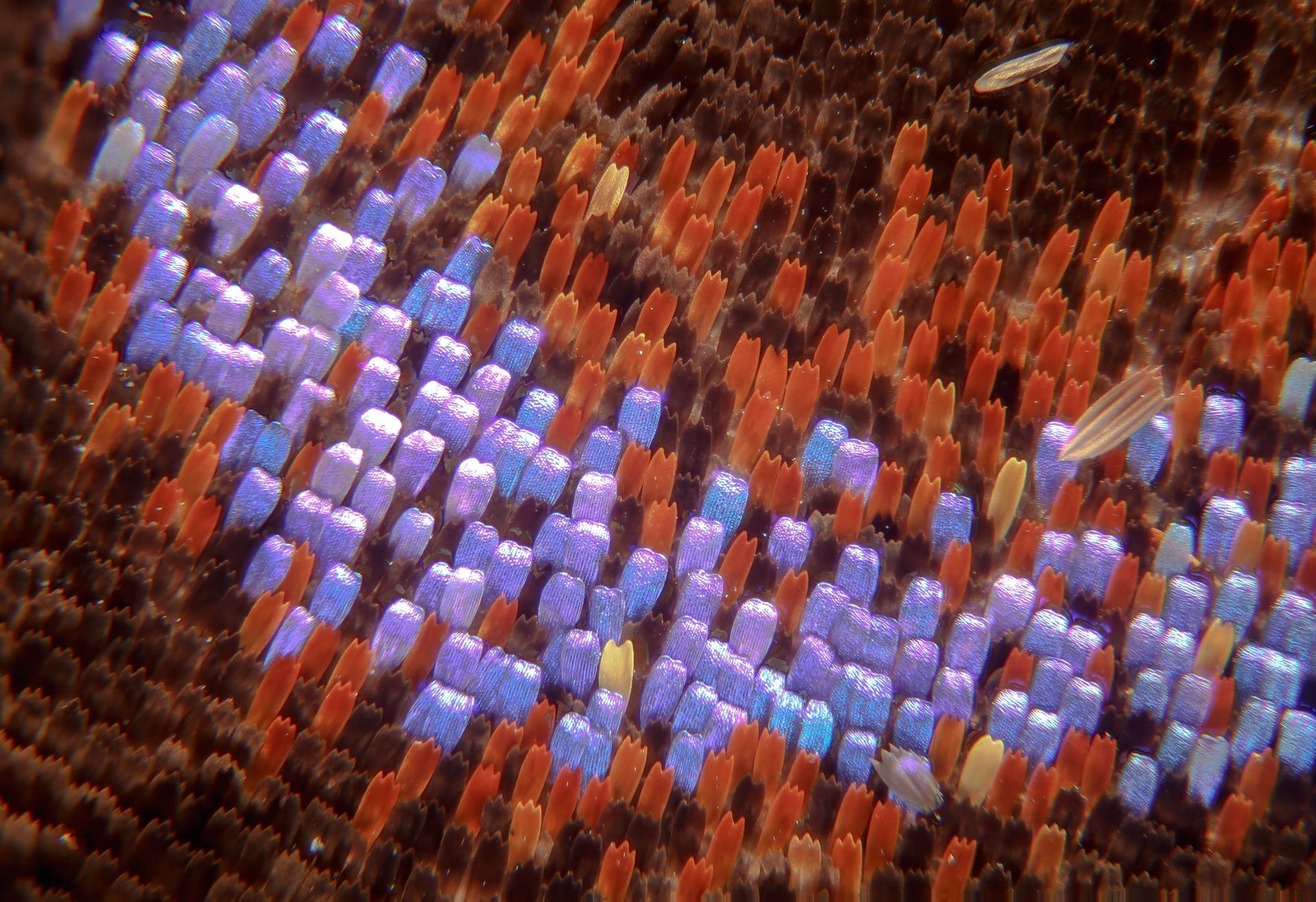
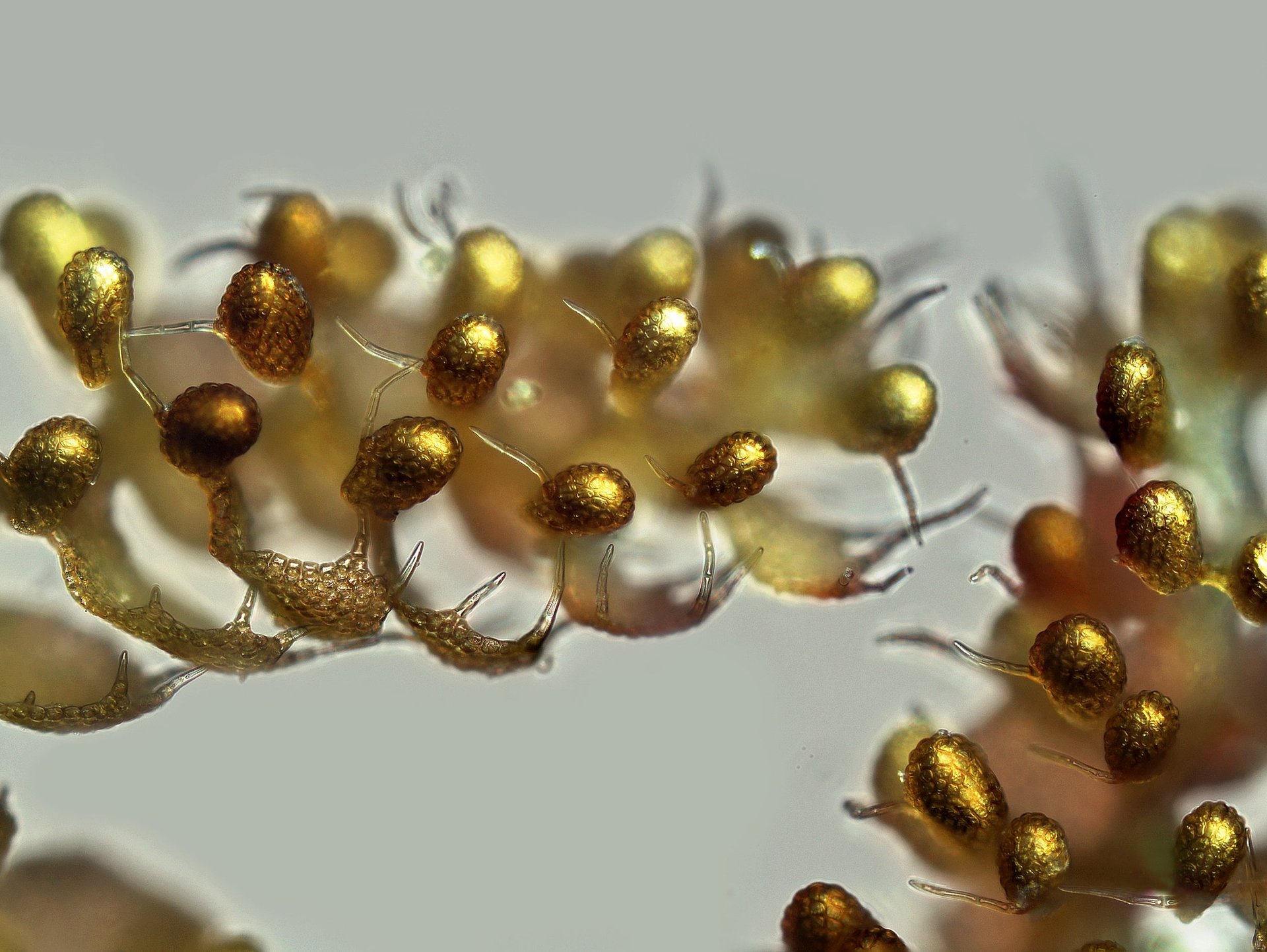
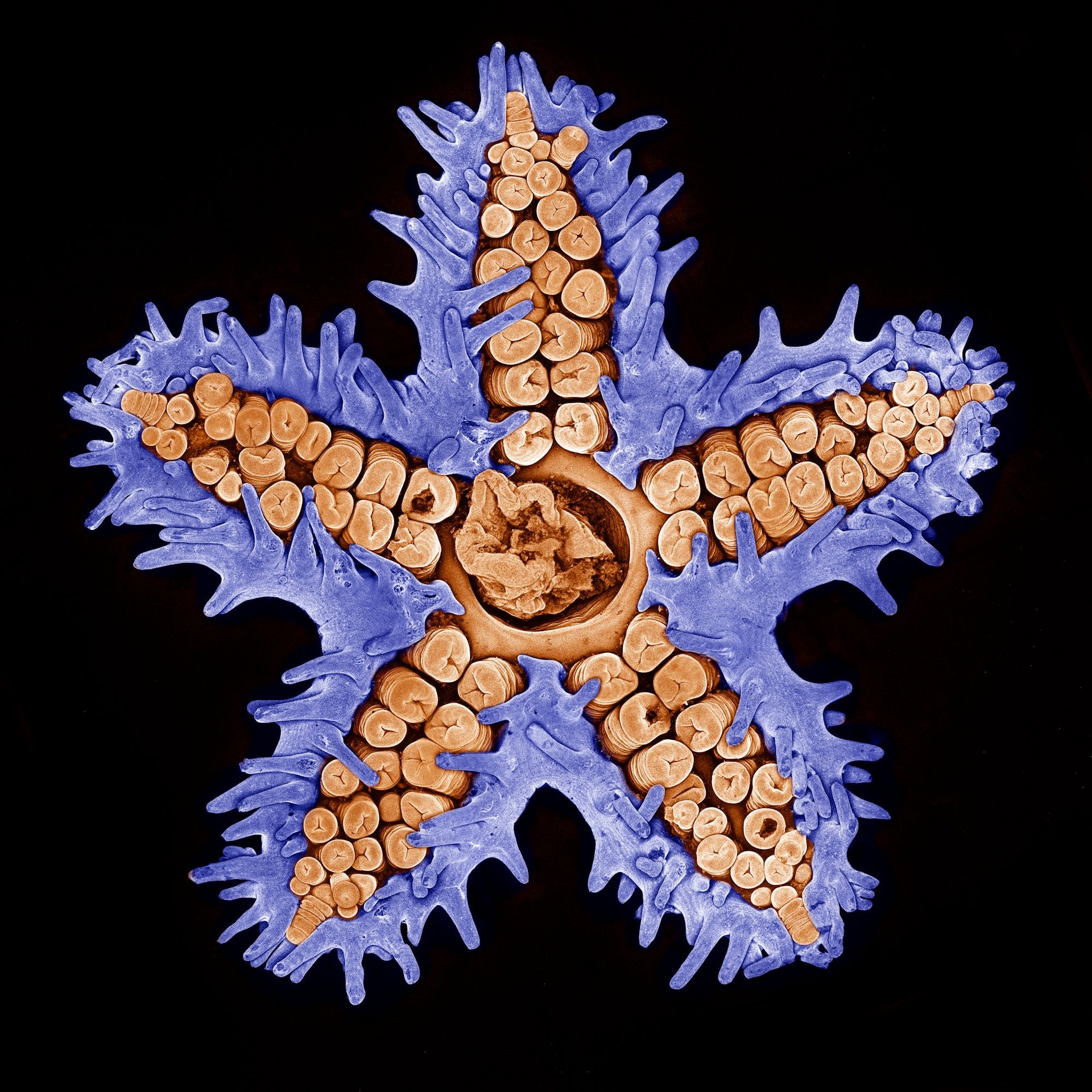
A few of the year’s top submissions:

Mouse colon colonized with human microbiota, magnified 63x.
Image: Kristen Earle, Gabriel Billings, KC Huang & Justin Sonnenburg/Stanford University School of Medicine, Department of Microbiology and Immunology/Nikon Small World
Adult marine worm, magnified 30x.
Image: Michael Crutchley/Nikon
Transverse section of an ostrich fern, magnified 250x.
Image: Anatoly Mikhaltsov/Nikon Small World
Tentacles of a carnivorous plant (Drosera sp.), magnified 20x.
Image: Jose Almodovar/University of Puerto Rico (UPR), Mayaguez Campus, Biology Department/Nikon Small World
Vilene fabric (glue drops shown in blue), magnified 80x.
Image: Gerd-A. Günther/Nikon Small World
Antenna of a male moth (Anisota sp.), magnified 100x.
Image: Dr. Igor Siwanowicz/Hughes Medical Institute (HHMI), Janelia Farm Research Campus, Leonardo Lab/Nikon Small World
Alona guttata (water flea), magnified 200x.
Image: Jacek Myslowski/Nikon Small World
Scales on moth wing, magnified 300x.
Image: Donald Parsons/Nikon Small World
Liverwort (Lepidolaena taylorii) plant showing modified leaves (water sacs), magnified 100x.
Image: Susan Tremblay/University of California, Berkeley/Nikon Small World
Red fossil coral slab, magnified 20x.
Image: Norm Barker/Johns Hopkins School of Medicine, Department of Pathology/Nikon Small World
Starfish imaged using confocal microscopy, magnified 10x.
Image: Evan Darling/Memorial Sloan Kettering Cancer Center /Nikon Small World