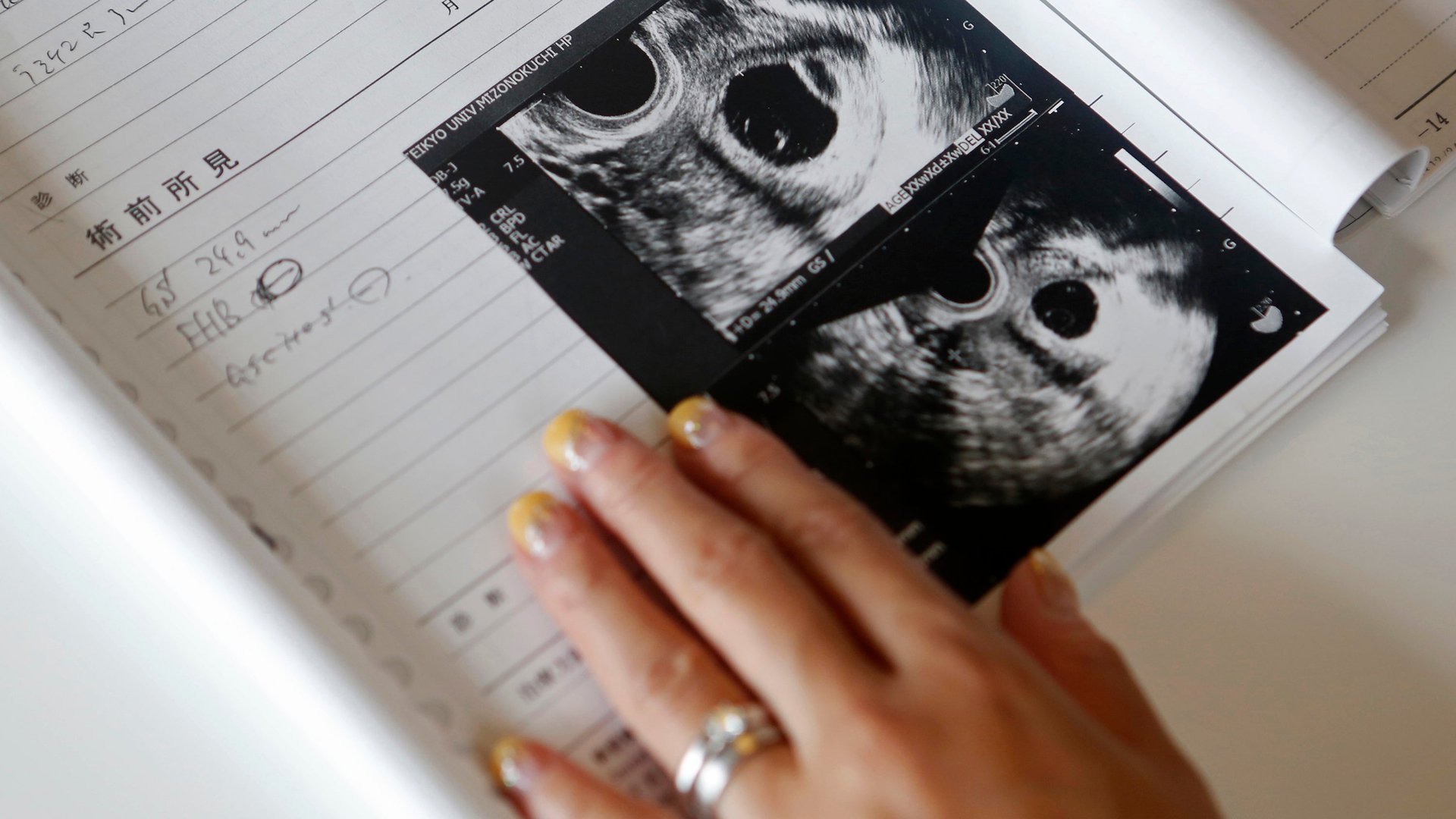
Welcome to the new 3D ultrasound economy
During my first pregnancy, I found myself gazing at a highly realistic 3D ultrasound of my unborn child. The level of detail was a far cry from the grainy, black-and-white blobs I was used to seeing. I could see the shape of my baby’s nose, the roundness of his cheeks, and a tiny, perfectly formed hand pressed against his forehead.


During my first pregnancy, I found myself gazing at a highly realistic 3D ultrasound of my unborn child. The level of detail was a far cry from the grainy, black-and-white blobs I was used to seeing. I could see the shape of my baby’s nose, the roundness of his cheeks, and a tiny, perfectly formed hand pressed against his forehead.
The advanced imagery turned out to be a lifesaver. Modern 3D and 4D ultrasound technology can be used to spot any number of birth defects and abnormalities in utero. In my case, the ultrasound found that my low-lying placenta had grown over my cervix, making a vaginal birth a danger to both me and my baby. Thanks to the test, I was able to deliver my son safely via Cesarean section.
But while technological advancements have made ultrasounds even more useful to expectant mothers, they’ve also made recreational ultrasounds increasingly popular–a practice that may expose fetuses to increased risks. Meanwhile, some women are raising concerns about the prospect of getting too much information about their unborn children. Here’s what the medical community has to say about responsible ultrasound use.
The ultrasound economy
Most pregnant women are familiar with 2D ultrasounds—the high-frequency sound waves that use echoes to create blurred, black-and-white images of a fetus. Used as a diagnostic tool, they can determine a baby’s due date, confirm the location of the placenta, measure amniotic fluid, and analyze things like fetal position, movement, and heart rate.
There’s also something breathtaking about the moment when you first catch sight of the little cashew-shaped creature taking up residence in your uterus. Radiology clinics regularly supply proud parents-to-be with images and DVDs of their frog-faced little fetuses. Now boutique clinics are capitalizing on popular demand, popping up in shopping malls to offer 3D/4D ultrasounds that aren’t medically necessary. They boast cute names like “Meet Your Baby” and “Baby’s Debut.”
These mementos have sparked concerns about the overuse of ultrasound technology. In 2014, the Federal Drug Administration (FDA) issued a statement warning mothers against fetal “keepsake” images, as well as the overuse of Doppler fetal ultrasound heartbeat monitors.
“Although ultrasound has a proven track record of safety, there exists the risk of potential harm to the fetus if this technology is misused,” Lyndsay Meyer, a spokeswoman for the FDA, wrote to me in an email.
According to the FDA, “fetal tissue may be heated slightly” during ultrasounds. We don’t yet understand the potential long-term effects of this heating. We similarly don’t know enough about “cavitation,” that is, the impact of ultrasounds on gas bubbles in living tissue.
Meanwhile, the spectral Doppler used in obstetrical examinations to detect blood flow can cause even more heating, especially in soft tissue, as it directs an ultrasound beam to one small area rather than being scanned across a larger field of view. This has led many medical societies to establish guidelines against using fetal Doppler early in pregnancies, since blood flow—which carries away generated heat—is not yet fully established in fetuses.
That said, medical research thus far has shown little to worry about. Though studies are limited, a 1993 trial compared women who received one ultrasound at 18 weeks with women who received ultrasound imaging and continuous-wave Doppler flow studies at 18, 24, 28, 34, and 38 weeks. The only difference between the two was that women with more frequent ultrasounds had significantly higher intrauterine growth restriction, or delayed growth, of the unborn baby. It was unclear whether this difference was a fluke or a direct result of the scans.
Still, in low-risk pregnancies, there’s no reason to expose fetuses to more ultrasounds than they need. The American College of Obstetricians and Gynecologists and the American College of Radiology both recommend no more than two ultrasounds in low-risk pregnancies.
“The ultrasounds we recommend are at 12-14 weeks looking for major defects and markers for Down Syndrome, and again at 18-20 weeks to do a full survey for abnormalities,” says Dr. Beryl Benacerraf, president of the American Institute of Ultrasound in Medicine and a clinical professor of radiology and obstetrics and gynecology at Harvard Medical School.
And yet, often times, women with low-risk pregnancies regularly receive between four and five ultrasounds—more than double the recommended amount, according to a 2013 study published in the journal Seminars in Perinatology.
Opting out
“What I see in my practice is not the risk of the ultrasound itself, but the risk of too much information,” said Becca Gordon, a Los Angeles-based doula who has attended more than 150 births in her career.
Gordon, who personally chose to have only one anatomical ultrasound when she was 23 weeks pregnant, said that she has seen many nebulous diagnoses in utero. While ultrasounds can spot potential conditions such as low amniotic fluid, delayed growth or large babies, those diagnoses aren’t always accurate. In such cases, expecting parents can experience unnecessary stress as well as medical interventions, such as inductions and C-sections, that they may have been able to do without.
False diagnoses in ultrasounds, according to Benacerraf, are not necessarily quantifiable: “The big problem with ultrasound technology is that it’s operator dependent. Unlike a chest X-ray image that someone else looks at later, the ultrasound is performed by a doctor or sonographer and the variability of skill and training is problematic.”
While there has been a movement to try to standardize training and accredit ultrasound practices, Benacerraf also explained, “Ultrasound is not perfect. It’s not going to diagnose everything and it’s going to find things that are not significant.”
My conversations about these “insignificant” diagnoses triggered a memory I had long put aside. A 3D ultrasound with my firstborn had detected a blockage that caused swelling in his right kidney. The conditioned resolved itself after his birth. But had he been born just one year earlier, medical protocol would have dictated that he immediately be put on antibiotics to avoid the risk of a kidney infection. Those antibiotics could have altered his gut bacteria, putting him at risk for illnesses later in life, including allergies and infectious diseases, according to a 2015 study in the journal Cell, Host and Microbe.
Of course, it’s not much use to fret over a scenario that never happened, for a medical condition that never fully manifested itself. But perhaps there are circumstances under which some women may feel they are better off not knowing.
Consider expecting mothers who know they would carry a baby to term no matter what—whether their baby has a genetic condition like Down Syndrome or a physical abnormality that could affect quality of life.
Benacerraf says that limiting ultrasounds to two in a low-risk pregnancy is “perfectly reasonable.” But she advises women against refusing prenatal screenings altogether.
“In my opinion, it’s short-sighted not to have a test because you’re afraid of the results,” she says. She explained, for example, that children with Down Syndrome can be vulnerable to other medical issues. So even if a mother intends to carry the child to term, it’s still critical to have that information in hand.
“If you choose to let nature take its course, maternal and fetal mortality can be very significant if you don’t avail yourself to modern medicine,” she said.
As for me and my current pregnancy, because of my fertility treatments and “advanced maternal age,” I’ve been shuffled into the high-risk group. That means I’m committed to riding out the ultrasound train which includes frequent 2D and 3D scans. I have to trust that the benefits will outweigh the risks—and I might as well admire my collection of colorful little baby scans. With childrearing, there’s always something to worry about. I’ll save my energy where I can.