
A closer look at how streaming services decide what to recommend, covering watch history, hidden microgenres, thumbnail tests and the algorithms behind your queue
Credit: National Institute of Allergy and Infectious Diseases / Unsplash
The immune system does not operate from a single command center. It runs as a distributed network of organs, cells and chemical signals spread from the skin to the bone marrow, and most of what it does happens without producing any symptom at all. A person can be exposed to thousands of microbes in a single day and never notice, because the system has already identified, contained and cleared the threat before it could cause harm. What people usually learn about immunity stops at a few familiar terms — white blood cells, antibodies, maybe T cells from a biology class. The actual system is built from dozens of specialized parts, each with a distinct job, and many of them work in ways that have nothing to do with the popular image of the body simply "fighting off germs."
Some of the most useful facts about immunity involve things that feel unrelated to disease at all: sleep quality, gut bacteria, aging, and stress hormones all directly change how well the immune system performs. Other facts explain symptoms that are often misread as problems, such as fever, which is not the body malfunctioning but the body deliberately raising its own temperature as a weapon. Understanding these mechanisms changes how a person interprets illness, vaccination, and even everyday habits like sleep and diet.
This list breaks down 20 specific, well-documented pieces of how the immune system actually functions, from the physical barriers that stop pathogens before they enter the body to the specialized cells that shut the whole response down once a threat is gone. Each one is a self-contained piece of the larger picture, drawn from established immunology rather than folk explanations. None of it requires a medical background to understand, and all of it explains something about the body that most people go their whole lives without learning.
Credit: CDC / Unsplash
The innate immune system responds to a threat within minutes to hours, using cells and proteins that recognize general patterns common to pathogens rather than a specific germ. This branch does not require prior exposure to work, which is why it is considered nonspecific.
Its main players are phagocytes, cells that physically engulf and digest invaders. Macrophages patrol tissues and consume bacteria, dead cells and debris, while neutrophils are usually first to arrive at a site of infection, drawn in by chemical signals released from damaged tissue. Both cell types carry pattern recognition receptors on their surface, molecular sensors built to detect structures found on many pathogens at once, such as bacterial cell wall components or viral genetic material.
Inflammation is the visible sign of this system at work. Redness, heat, swelling and pain around a wound or infection come from blood vessels widening and becoming more permeable, allowing immune cells and fluid to flood the area. This is a controlled, purposeful process, not simply damage.
Because innate immunity does not build a memory of specific pathogens, it responds the same way every time it encounters a threat, whether that threat is brand new or one the body has faced a hundred times before. That job — remembering — belongs to the adaptive immune system, which works alongside innate immunity but takes days rather than hours to fully activate. The two systems communicate constantly: innate immune cells often trigger and shape the adaptive response that follows, handing off information about what they encountered so a more targeted response can follow.

Credit: cottonbro studio / Pexels
A fever is the body raising its own internal temperature on purpose, as a calculated defense rather than a sign that something has gone wrong. The hypothalamus, a small region at the base of the brain, controls core body temperature and resets it upward in response to chemical signals called pyrogens.
These pyrogens include cytokines such as interleukin-1, interleukin-6 and tumor necrosis factor-alpha, released by immune cells after they detect an infection. When these signals reach the hypothalamus, it shifts the body's internal thermostat to a higher setpoint, and the body responds by generating heat through shivering and reducing heat loss by narrowing blood vessels near the skin. That combination is why a person with a rising fever often feels cold and shivers even as their internal temperature climbs.
The higher temperature works in two ways. Many bacteria and viruses replicate less efficiently at temperatures above the human baseline of about 98.6 degrees Fahrenheit, so raising body temperature by even a few degrees can slow a pathogen down directly. At the same time, several immune functions speed up in warmer conditions, including the migration of white blood cells to infection sites and the efficiency of some antibody responses.
Fever is capped by the body's own regulatory systems, which is why healthy adults rarely develop temperatures high enough to cause harm on their own, though very high or prolonged fevers do warrant medical attention, particularly in infants and young children. Suppressing a mild fever with medication does not stop the underlying immune response, since the innate and adaptive systems are already working through other pathways; fever is one tool among several, not the entire response, and lowering it is generally about comfort rather than altering the outcome of the illness itself.
Credit: Monstera Production / Pexels
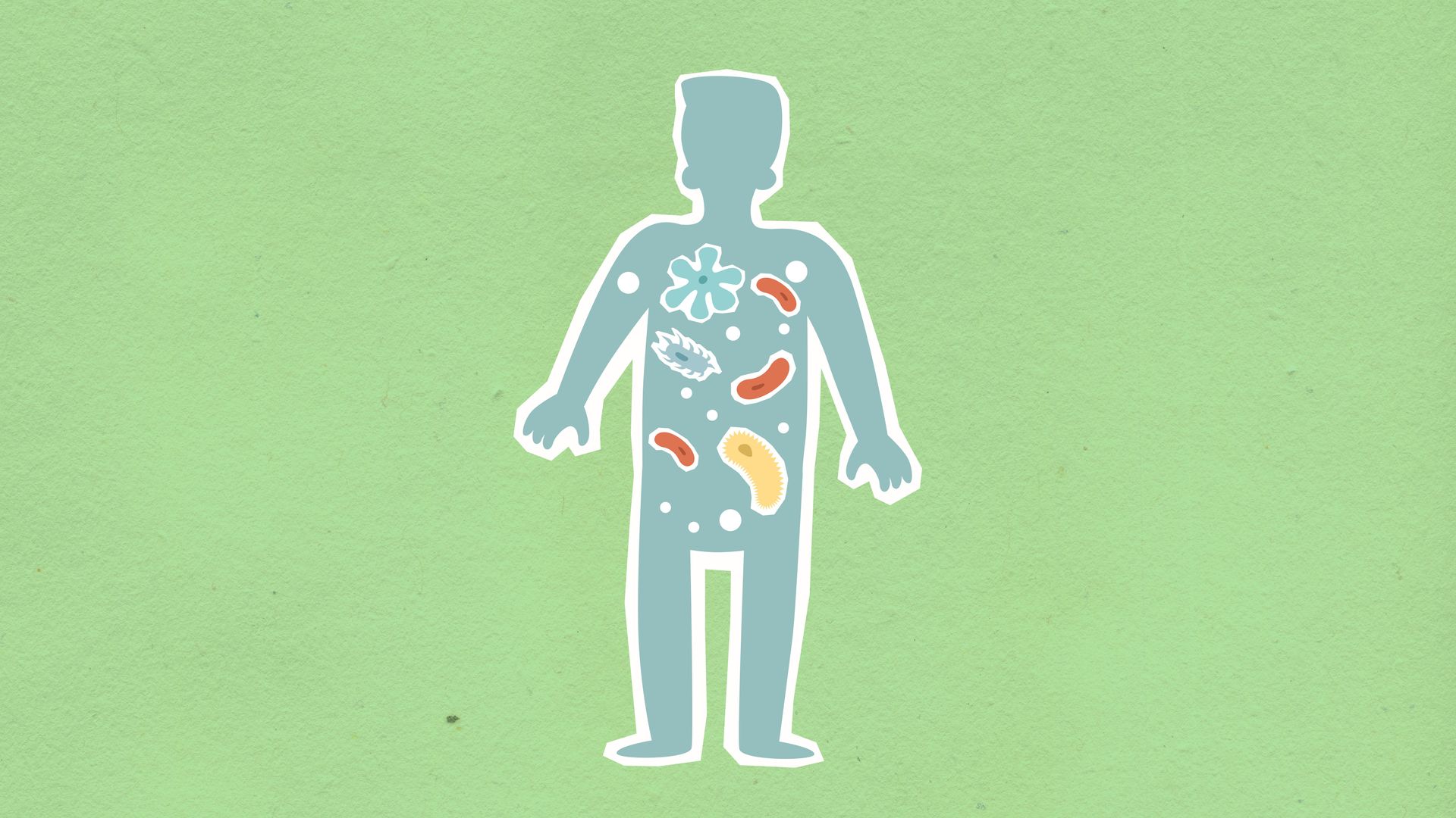
The trillions of bacteria living in the human gut do more than aid digestion; they directly train the immune system, particularly during infancy and early childhood. Roughly 70 to 80 percent of the body's immune cells are located in tissue surrounding the gut, called gut-associated lymphoid tissue, placing the immune system in constant contact with this bacterial population.
Early exposure to a diverse range of gut bacteria appears to teach the developing immune system which signals represent harmless residents and which represent genuine threats. Children raised in environments with limited microbial exposure show, in some studies, higher rates of immune-related conditions later in life, a pattern that has shaped ongoing research into the relationship between early microbial exposure and immune regulation.
Gut bacteria also produce byproducts that directly influence immune cell behavior. When bacteria ferment dietary fiber, they generate short-chain fatty acids, including one called butyrate, which supports the development of regulatory T cells, a specialized cell type that helps keep the immune system from overreacting to harmless substances. A gut community low in fiber-fermenting bacteria produces less of this signal, which some research links to a less well-regulated immune response.
The relationship runs in both directions. The immune system, in turn, helps keep the gut bacterial population in balance, using antibodies and antimicrobial proteins secreted into the gut lining to prevent any single bacterial species from overgrowing. This constant back-and-forth between gut bacteria and immune tissue is one reason antibiotics, which disrupt the microbiome broadly, can have effects on immune function that last well beyond the course of treatment, and it is part of why diet and gut health are now studied as direct levers on immune performance rather than separate systems entirely.
Credit: Arizona Science Center / Wikimedia Commons (CC BY-SA 3.0)
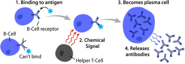
Memory B cells are the reason a person who recovers from an infection, or receives a vaccine, often has lasting protection against that specific pathogen. After an initial exposure, a subset of activated B cells does not die off once the infection clears; instead, they convert into long-lived memory cells that can persist in the bone marrow and lymph nodes for years, sometimes for life.
These memory cells carry a molecular record of the exact pathogen they encountered, in the form of a B cell receptor shaped to recognize a specific antigen. If that same pathogen appears again, memory B cells activate far faster than the original response required, often producing large quantities of targeted antibodies within days rather than the one to two weeks a first-time infection typically takes to control.
This mechanism is the biological basis for both natural immunity after infection and vaccine-induced immunity. A vaccine works by presenting the immune system with a harmless version of a pathogen's antigen, prompting the same memory B cell formation without requiring a person to go through the disease itself. Some vaccines produce memory that lasts decades, such as the measles vaccine, while others fade faster, which is part of why certain vaccines require booster doses.
Not every pathogen produces equally durable memory. Viruses that mutate quickly, such as influenza and the viruses that cause the common cold, present a moving target, since memory B cells built around one strain's antigen may not recognize a mutated version well. This is a central reason flu vaccines are reformulated most years, and why a person can catch a cold multiple times in one season despite an intact and fully functional memory B cell system.
Credit: CDC / Pexels
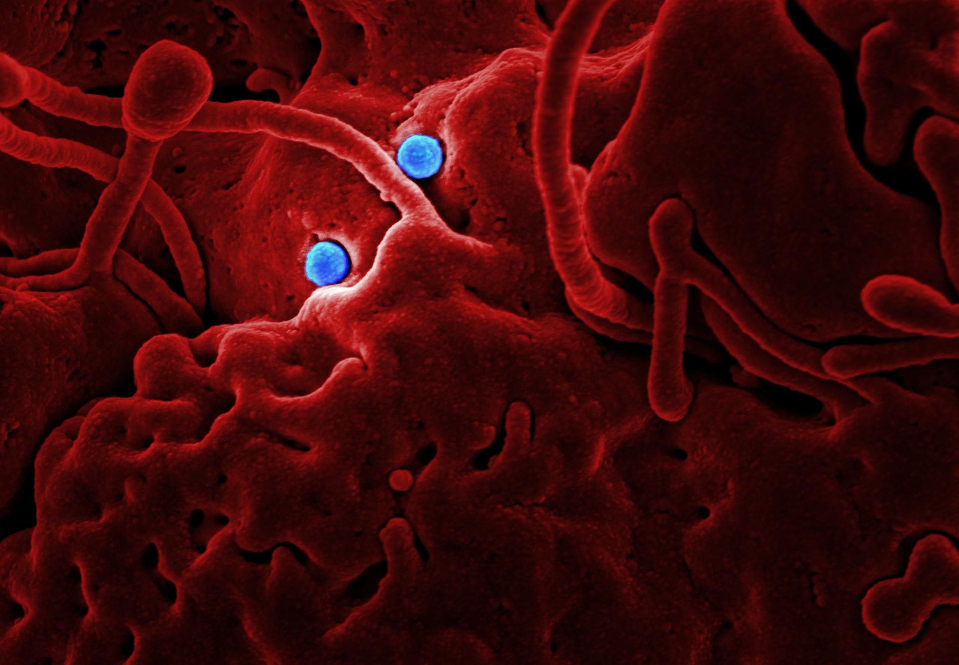
Natural killer cells, or NK cells, are a type of white blood cell that can destroy virus-infected and cancerous cells without needing to recognize a specific antigen first, setting them apart from most of the adaptive immune system. They are technically part of the innate branch, despite being a type of lymphocyte, the same broad cell family that includes T cells and B cells.
Most cells in the body display a marker on their surface called MHC class I, a kind of identification tag that signals to the immune system that the cell is healthy and belongs to the body. NK cells are built to detect the absence of this marker. Many viruses and cancerous cells suppress MHC class I display as a way to hide from T cells, but that same suppression exposes them to NK cells, which are tuned to attack exactly the cells that look like they are hiding something. This is often called the "missing self" mechanism.
Once an NK cell identifies a target, it releases proteins called perforin and granzymes directly onto the target cell's surface. Perforin creates small pores in the cell membrane, and granzymes pass through those pores to trigger a controlled cell death process inside the target, destroying it from within without damaging surrounding healthy tissue.
NK cells also release cytokines, including interferon-gamma, that alert and recruit other immune cells to the area, functioning as an early warning system as well as a direct attacker. Because they act without needing prior exposure or a slow activation process, NK cells provide a rapid response against infected or abnormal cells in the earliest hours of an immune reaction, well before T cells have been activated and multiplied in sufficient numbers to join the fight.
Credit: OpenStax College - Anatomy & Physiology, Connexions Website / Wikimedia Commons (CC BY 3.0)
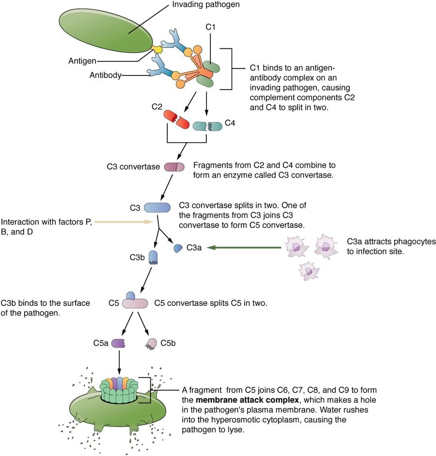
The complement system is a group of about 30 proteins that circulate in the blood in an inactive form and activate in a chain reaction once triggered, rather than through a single dedicated cell type. It is one of the oldest and most direct weapons the immune system has against bacteria.
Complement proteins can be triggered through three separate pathways: one activated by antibodies already bound to a pathogen, one triggered directly by molecules on a pathogen's surface, and one that activates spontaneously at a low background rate and gets amplified once it detects a foreign surface. All three pathways converge on the same outcome, forming what is called a membrane attack complex, a ring-shaped structure that inserts into a bacterial cell's outer membrane and punches an actual hole through it. Fluid rushes into the cell through that hole and the bacterium bursts.
Complement proteins have other jobs beyond direct killing. Some coat, or opsonize, the surface of pathogens, effectively tagging them so that phagocytes such as macrophages recognize and consume them more efficiently. Others, called anaphylatoxins, are released during the cascade and act as chemical signals that recruit more immune cells to the site and increase local blood vessel permeability, contributing directly to inflammation.
Because the complement cascade can escalate quickly and cause tissue damage if it runs unchecked, the body carries several regulatory proteins whose entire job is to prevent complement from activating against the body's own healthy cells. Genetic deficiencies in these regulatory proteins are linked to certain autoimmune and inflammatory conditions, illustrating how a defense mechanism built for speed and force also requires precise internal controls to avoid becoming a liability to the same body it protects.
Credit: National Institute of Allergy and Infectious Diseases / Unsplash
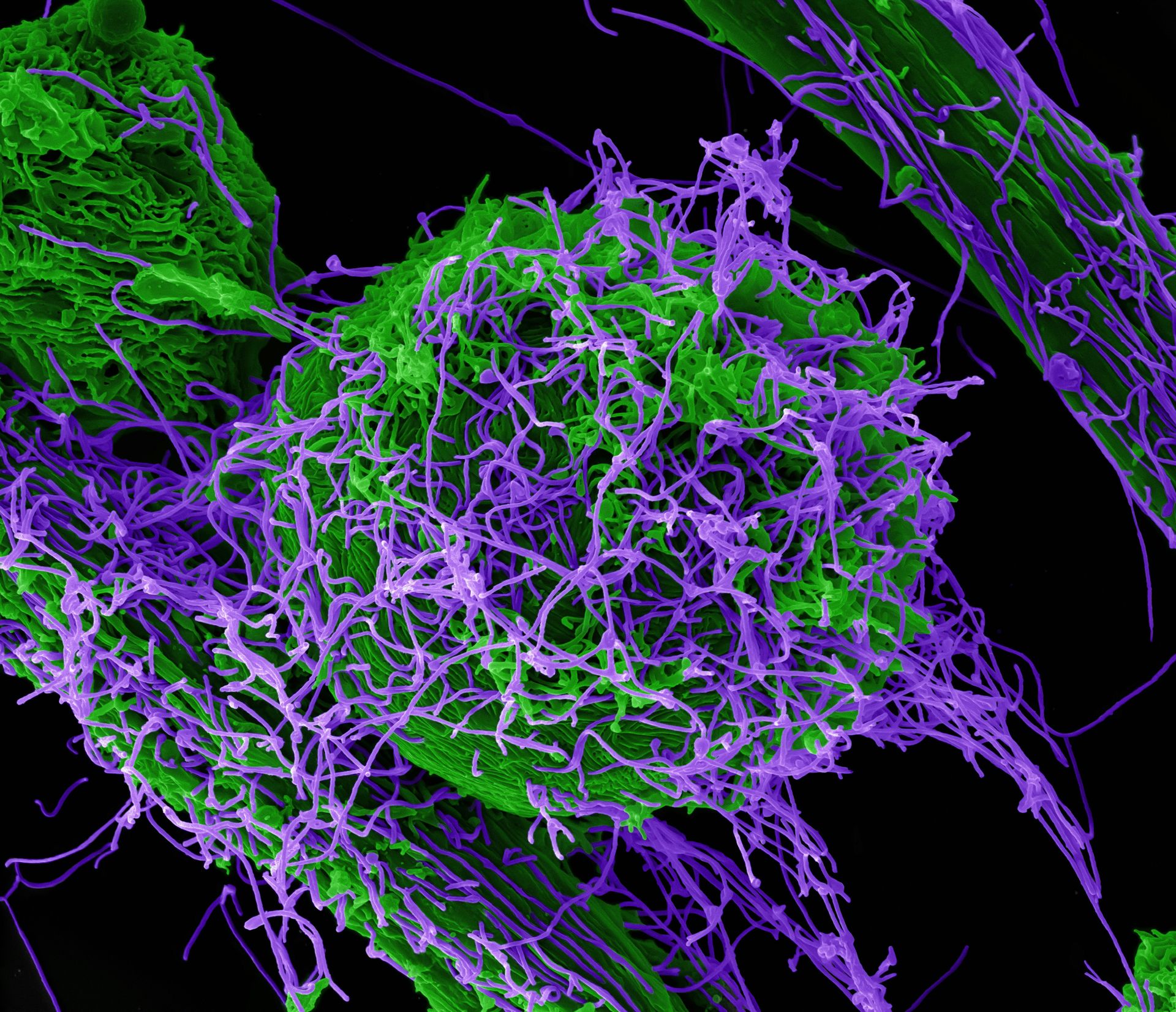
Neutrophils, the most abundant type of white blood cell, have a defense mechanism beyond simply engulfing pathogens: they can release their own DNA in a web-like structure that traps and kills microbes outside the cell. These structures are called neutrophil extracellular traps, or NETs, and their role in immunity was first described in detail in 2004.
To form a NET, a neutrophil breaks down its own internal nuclear membrane, releasing chromatin, the combination of DNA and proteins normally packed tightly inside the cell's nucleus. That chromatin, studded with antimicrobial proteins and enzymes such as elastase and myeloperoxidase, is expelled outward in a sticky, fibrous mesh. Bacteria and fungi become physically caught in this mesh, where the attached antimicrobial proteins can kill them or at least stop them from spreading further through tissue.
This process, sometimes described as a form of cell death distinct from ordinary programmed cell death, effectively sacrifices the neutrophil itself in exchange for containing an infection that might otherwise spread faster than individual cells could consume it. NETs are particularly useful against pathogens too large for a single neutrophil to engulf, or against fast-multiplying bacteria in situations where speed of containment matters more than a clean kill.
NETs are not without cost to the body. The same DNA and proteins that trap pathogens can contribute to tissue damage and blood clotting when released in large quantities, and excessive NET formation has been linked to conditions including certain autoimmune diseases and complications seen in severe infections. Researchers continue to study how to harness the pathogen-trapping benefit of NETs while limiting the collateral damage they can cause in the surrounding tissue when the response is prolonged or excessive.
Credit: OpenStax College - Anatomy & Physiology, Connexions Website / Wikimedia Commons (CC BY 3.0)
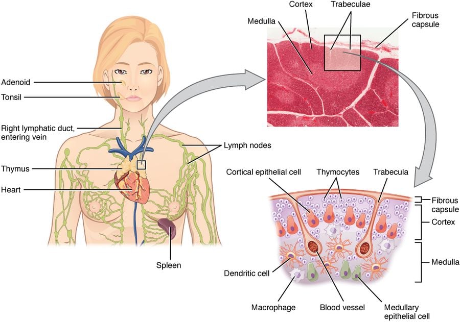
The thymus, a small gland located behind the breastbone, is where T cells mature and learn to distinguish the body's own cells from foreign material. Unlike most organs, it reaches its largest size in childhood and then steadily shrinks, in a well-documented process called thymic involution.
Immature T cells originate in the bone marrow but travel to the thymus to complete their development. Inside the thymus, each developing T cell is tested against a wide range of the body's own proteins in a process of positive and negative selection: cells that fail to recognize the body's own tissue-compatibility markers are eliminated, and cells that react too strongly against the body's own proteins are also eliminated, leaving behind a population of T cells capable of recognizing foreign threats without attacking healthy tissue.
Thymic tissue is at its most active before puberty. After that point, functional thymic tissue is gradually replaced by fatty tissue, and by middle age, only a small fraction of the gland's peak capacity remains. This means the rate at which the body can generate brand-new, "naive" T cells — ones that have never encountered a specific pathogen before — declines steadily over a person's lifetime.
This decline is one reason older adults often mount weaker responses to new pathogens and to new vaccines, since the body relies increasingly on existing memory T cells rather than generating fresh ones suited to a novel threat. It does not mean older immune systems stop working; memory T cells built earlier in life, along with B cells and innate immunity, continue functioning normally. The specific vulnerability is to genuinely new threats the body has not encountered before, which is part of why vaccination in childhood, while thymic output is still high, has an outsized long-term effect on lifetime immunity.
Credit: National Institute of Allergy and Infectious Diseases / Unsplash
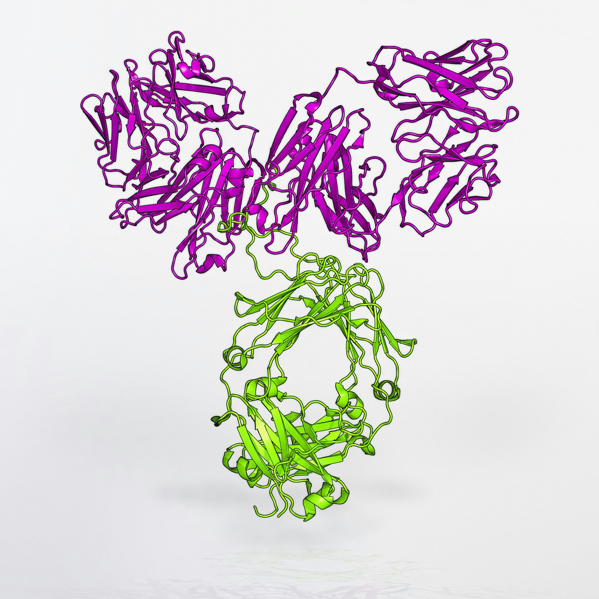
Antibodies, also called immunoglobulins, are not a single uniform tool but come in five structurally distinct classes, each suited to a different location in the body or stage of an immune response. These classes are labeled IgG, IgM, IgA, IgD and IgE.
IgG is the most abundant antibody in the bloodstream and the main long-term antibody produced after infection or vaccination. It is small enough to cross the placenta, giving a developing fetus passive immunity from the mother that lasts for the first few months of life. IgM is the first antibody type produced during a new infection, appearing within days; it forms a large five-unit structure that makes it especially effective at triggering the complement system, though it fades as IgG production ramps up.
IgA is the dominant antibody at mucosal surfaces, found in saliva, tears, breast milk and the linings of the respiratory and digestive tracts, where it intercepts pathogens before they can penetrate deeper into tissue. IgE, while present in the bloodstream in very small amounts under normal conditions, is best known for its role in allergic reactions, binding to mast cells and basophils and triggering the release of histamine when it encounters an allergen such as pollen or a food protein. IgE also plays a defensive role against parasitic worm infections, a function that predates its more commonly discussed role in allergies.
IgD is the least understood of the five classes. It is found mainly on the surface of immature B cells, where it appears to function as a receptor that helps regulate how those B cells respond to antigens before fully maturing, though its precise role remains an active area of immunology research compared with the other four, better-characterized classes.
Credit: Henry Gray, Anatomy of the Human Body (1918) / GetArchive
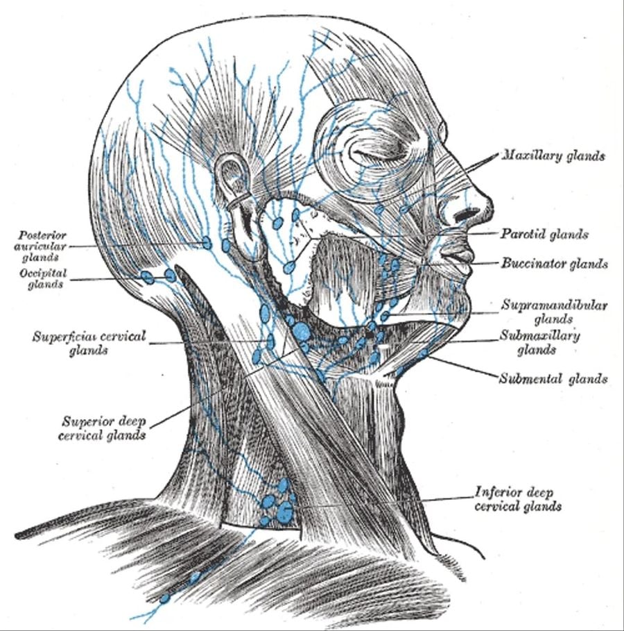
The lymphatic system is a separate network of vessels running parallel to the circulatory system, and it functions as the immune system's primary transportation and surveillance network. Rather than carrying blood, it carries a clear fluid called lymph, which picks up excess fluid, proteins and immune cells from tissues throughout the body.
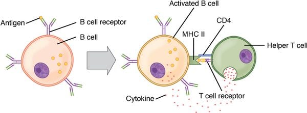
Lymph nodes, small bean-shaped structures scattered along this network, act as checkpoints where immune cells congregate and filter the fluid passing through. Lymph nodes are densely packed with lymphocytes, including B cells and T cells, along with dendritic cells that display fragments of pathogens they have encountered elsewhere in the body. When a dendritic cell carrying a piece of a pathogen arrives at a lymph node, it can activate specific T cells and B cells that recognize that exact antigen, kicking off a targeted adaptive immune response.
This explains why lymph nodes near an infection often swell and become tender, a sign that the nodes are filling with activated immune cells actively working through a response. A swollen lymph node under the jaw during a throat infection, for example, reflects the node's role as a local staging ground rather than a site of infection itself.
Beyond transporting immune cells, the lymphatic system also returns excess fluid from tissues back into the bloodstream, preventing fluid buildup, and it absorbs fats from the digestive tract through specialized vessels in the small intestine. The spleen, tonsils and adenoids are also considered part of the broader lymphatic system, each contributing additional sites where immune cells gather, monitor for threats and mount responses, making the lymphatic network less a single organ and more a distributed surveillance system running throughout nearly every region of the body.
_diagram-1920x1261.png)
Credit: A. Rad / Wikimedia Commons (CC BY-SA 3.0)
Before any immune cell has to respond to a pathogen, the body's physical barriers are doing most of the work, and skin is the largest of them. The outermost layer of skin, called the stratum corneum, is made of flattened, dead, keratin-filled cells packed tightly together, forming a barrier most microbes cannot penetrate on their own.
Skin also maintains a slightly acidic surface, often called the acid mantle, created by sweat, oil and the natural bacteria that live on skin, and this acidity discourages the growth of many harmful microorganisms. Skin cells additionally secrete antimicrobial peptides, small proteins capable of directly damaging bacterial cell membranes on contact, adding a chemical defense layer to the physical one.
Mucous membranes, lining the respiratory tract, digestive tract and other internal surfaces exposed to the outside world, use a different strategy built around a thick, sticky substance called mucus. Mucus physically traps inhaled particles, bacteria and viruses before they can reach deeper tissue. In the airway, tiny hair-like structures called cilia beat in a coordinated wave, sweeping mucus and everything trapped in it upward and out of the lungs, a process known as the mucociliary escalator.
Additional chemical defenses reinforce these barriers at specific sites. Tears and saliva contain an enzyme called lysozyme, which breaks down the cell walls of many bacteria on contact, and stomach acid is potent enough to destroy most microbes swallowed with food or water. Together, these barriers represent the first stage of defense the innate and adaptive immune systems never have to get involved in, since most microbial exposures are neutralized here before they ever penetrate far enough to trigger a full immune response.
Credit: Sonu Bhaskar, Akansha Sinha, Maciej Banach, Shikha Mittoo, Robert Weissert, Joseph S. Kass, Santhosh / Wikimedia Commons (CC BY 4.0)
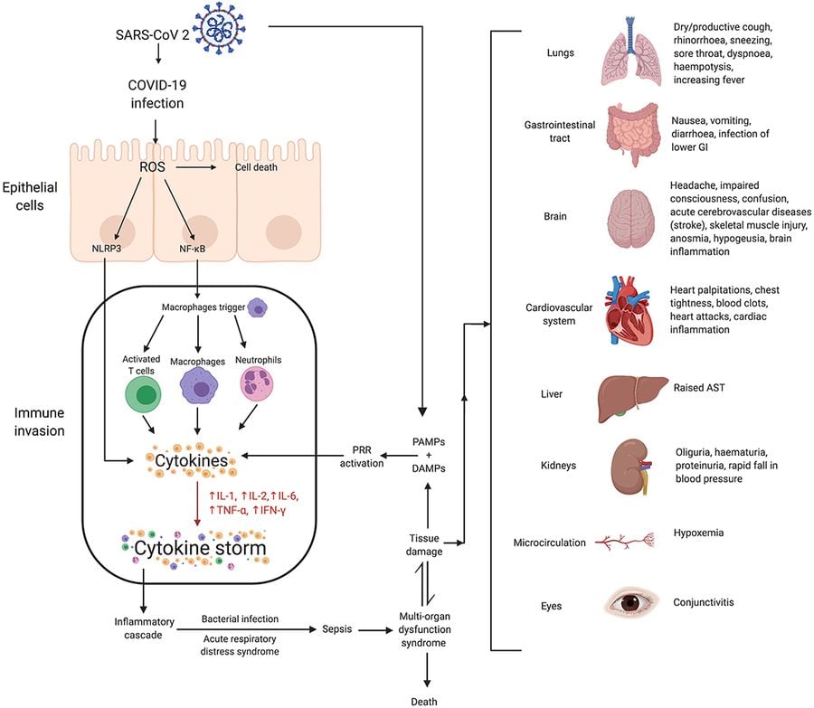
A cytokine storm occurs when the immune system releases inflammatory signaling proteins, called cytokines, in such large and sustained quantities that the response itself begins damaging the body's own tissue, rather than only the pathogen it was meant to fight. It represents a breakdown of the normal checks that keep an immune response proportional to the threat.
Cytokines such as interleukin-6 and tumor necrosis factor-alpha are normally released in controlled amounts to recruit immune cells, trigger fever and coordinate a response. In a cytokine storm, feedback loops that would typically dial this signaling back instead amplify it, with activated immune cells producing more cytokines that recruit and activate still more immune cells, in a cycle that can escalate rapidly over hours.
The effects extend well beyond the original site of infection. Circulating through the bloodstream, excess cytokines can cause blood vessels to become abnormally permeable, blood pressure to drop, and organs including the lungs, kidneys and liver to sustain direct damage from the inflammatory response itself rather than from the pathogen. This is part of why some severe infections, including certain cases of sepsis and severe cases of COVID-19, cause organ damage that appears disproportionate to the infection's initial severity.
Cytokine storms are not limited to infections. They have also been documented as a side effect of certain cancer immunotherapies, particularly CAR T-cell therapy, where immune cells engineered to attack cancer cells trigger a similarly rapid, large-scale cytokine release. Recognizing this risk, doctors administering these therapies now monitor patients closely for early signs of cytokine storm and have specific treatments, including drugs that block IL-6 signaling, ready to interrupt the cycle if it begins.

Credit: Ali Rowshani / Unsplash
Autoimmune disease occurs when the immune system fails to distinguish the body's own tissue from foreign material and mounts an attack against healthy cells, using the same tools it would normally direct at pathogens. More than 80 distinct autoimmune conditions have been identified, each targeting different tissues.
Normally, the immune system develops tolerance to the body's own proteins through two overlapping processes. Central tolerance happens in the thymus and bone marrow, where developing T cells and B cells that react too strongly to the body's own proteins are eliminated before they ever enter circulation. Peripheral tolerance acts as a backup, using regulatory T cells and other mechanisms throughout the body to suppress any self-reactive immune cells that slipped through the first filter. Autoimmune disease develops when both layers of tolerance fail for a specific target tissue.
The tissue targeted determines the disease and its symptoms. In type 1 diabetes, the immune system attacks insulin-producing cells in the pancreas. In rheumatoid arthritis, it targets the lining of joints, causing chronic inflammation and joint damage. In multiple sclerosis, it attacks the protective coating around nerve fibers in the brain and spinal cord. In Hashimoto's thyroiditis, it targets the thyroid gland, gradually reducing its ability to produce hormones.
The exact triggers that cause tolerance to break down remain an active area of research, but genetics, certain infections, and environmental factors are all understood to play a role in different combinations depending on the specific disease. Autoimmune conditions are also more common in women than men for reasons that are still being investigated, with hormonal and genetic factors both considered likely contributors to that pattern.
Credit: https://www.scientificanimations.com / Wikimedia Commons (CC BY-SA 4.0)
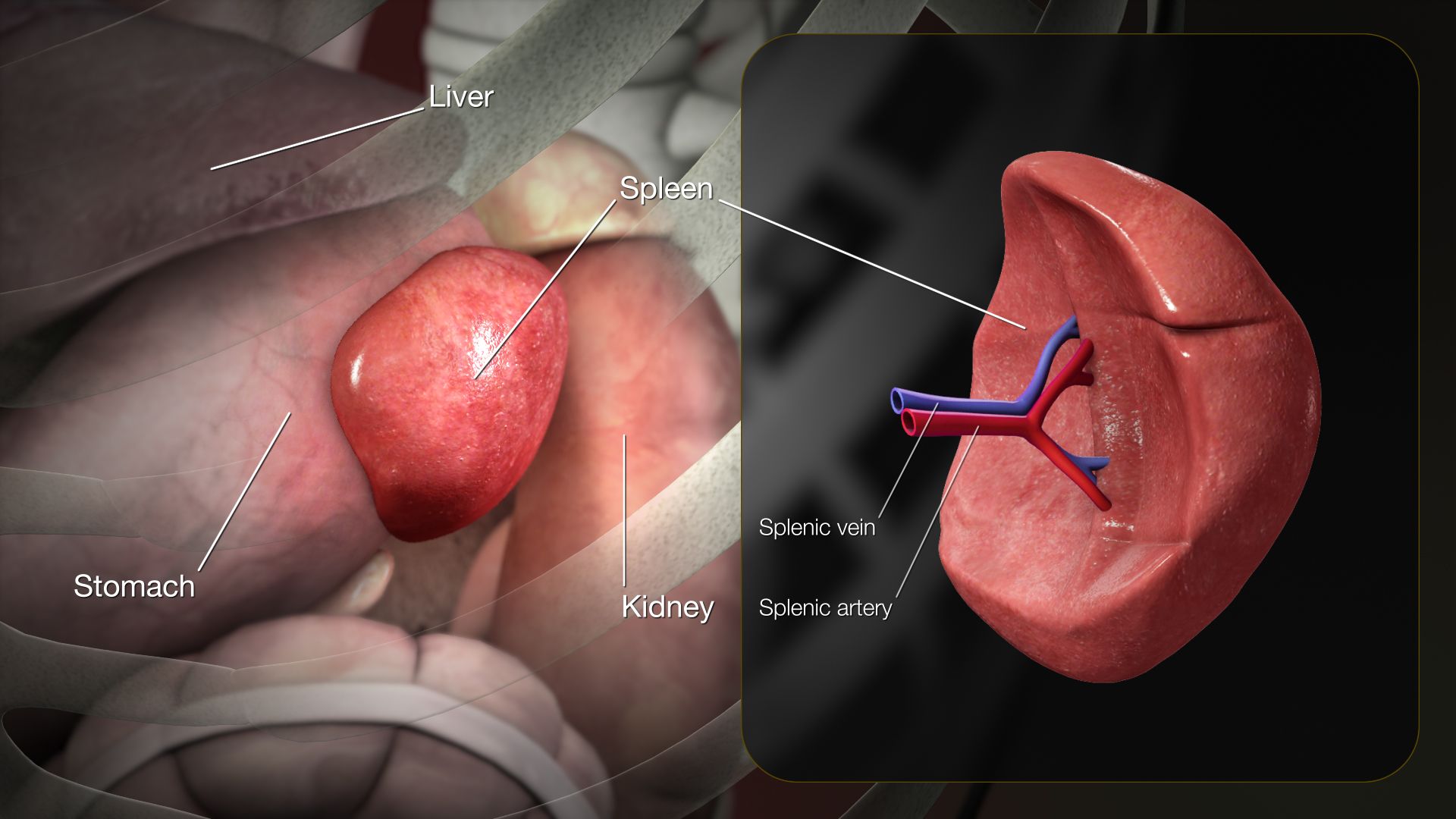
The spleen is the largest lymphoid organ in the body, located in the upper left abdomen, and it performs two distinct jobs: filtering blood and supporting immune responses. It is divided into two functional regions known as red pulp and white pulp, each handling a different task.
Red pulp is where the spleen filters blood directly, identifying and removing old, damaged or misshapen red blood cells from circulation. Red blood cells typically live about 120 days, and as they age and become less flexible, the spleen's red pulp catches and breaks them down, recycling iron from their hemoglobin for reuse elsewhere in the body, primarily in the production of new red blood cells.
White pulp is where the spleen contributes directly to immune defense. It contains dense clusters of lymphocytes, similar to the tissue found in lymph nodes, and it mounts immune responses specifically against pathogens present in the bloodstream, a role lymph nodes are not positioned to handle since they filter lymph rather than blood directly. The spleen also stores a reserve supply of monocytes, a type of white blood cell that can be rapidly deployed to a site of injury or infection when needed.
Because the spleen plays this dual role, its removal, whether due to injury or disease, leaves a person more vulnerable to certain infections, particularly from encapsulated bacteria such as Streptococcus pneumoniae, which the spleen is normally well equipped to filter from the blood and target with antibodies. People without a spleen typically receive additional vaccinations and, in some cases, preventive antibiotics to compensate for this specific gap in their immune defenses, since no other single organ fully replaces the spleen's combined filtering and immune functions.
Credit: FRANK MERIÑO / Pexels
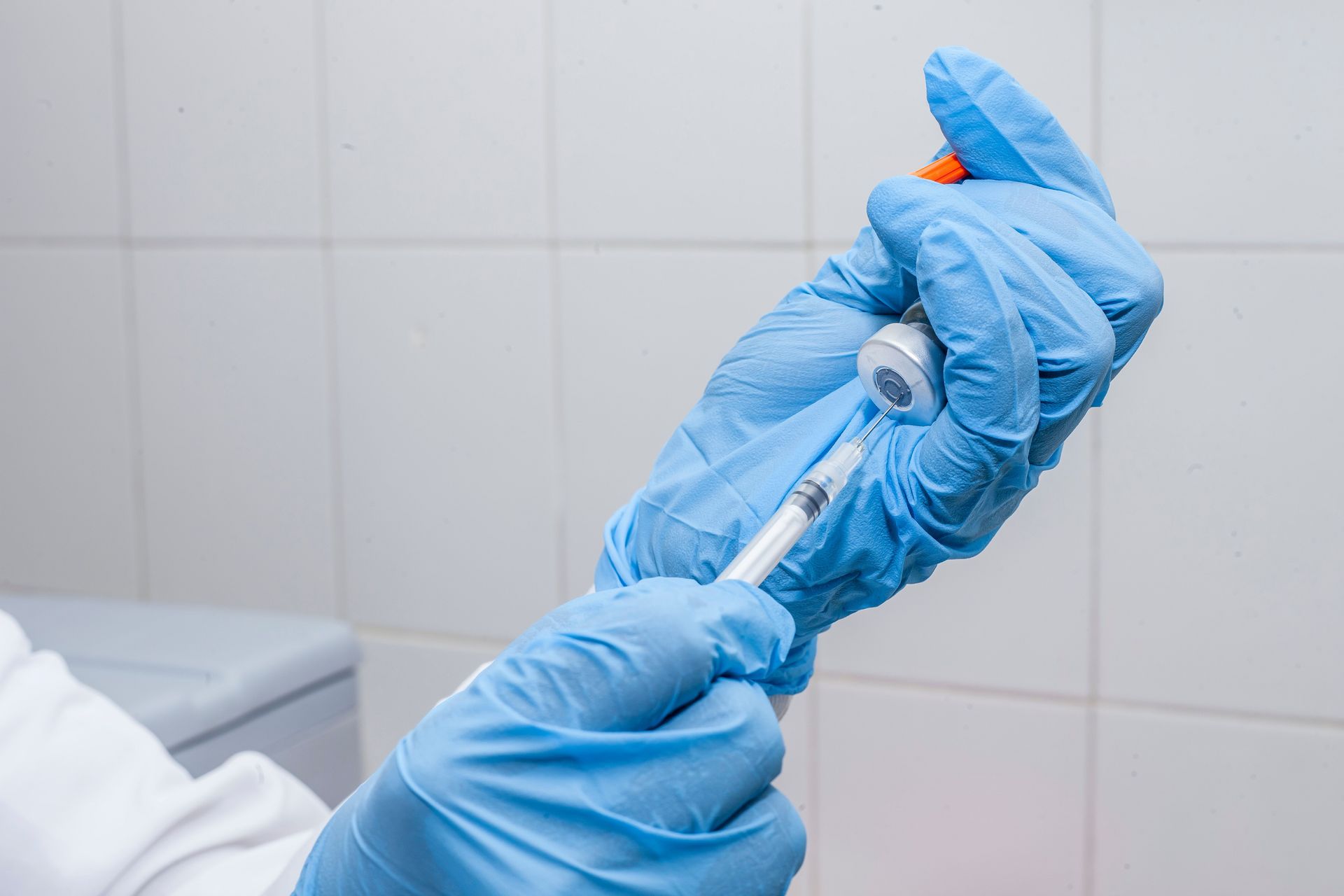
A vaccine works by introducing the immune system to a harmless version of a pathogen's antigen, prompting it to build the same memory B cells and T cells it would develop from a natural infection, without requiring a person to experience the actual disease or its complications.
Different vaccine types achieve this in different ways. Live attenuated vaccines, such as the measles, mumps and rubella vaccine, use a weakened form of the actual virus that can still replicate briefly but does not cause serious illness in a healthy person, generating a particularly strong and long-lasting immune response. Inactivated vaccines, such as most flu shots, use a killed version of the pathogen that cannot replicate at all. Subunit vaccines, including the hepatitis B vaccine, use only a specific piece of the pathogen, such as a single protein, rather than the whole organism. Messenger RNA vaccines, a newer category that includes some COVID-19 vaccines, instead deliver genetic instructions that prompt a person's own cells to temporarily produce a harmless piece of the pathogen's protein, which the immune system then recognizes and responds to.
Regardless of the method, the goal is the same: expose the immune system to a recognizable piece of the pathogen so it builds memory cells capable of responding quickly if the real pathogen is ever encountered. This is why a vaccinated person who is later exposed to the actual disease typically experiences a much faster, stronger response than someone with no prior exposure, often stopping the infection before symptoms develop at all.
Not every vaccine produces equally durable protection, which is part of why some, including tetanus and certain pertussis vaccines, require periodic booster doses to maintain strong memory cell populations over time, while others generate memory that lasts for decades with a single series of doses.

Credit: Polina / Pexels
Sleep is not simply rest for the brain; it directly affects how well the immune system functions, with measurable effects on infection risk and vaccine response. Several distinct mechanisms link the two systems.
During deep sleep, T cells show increased activity of a type of surface protein called an integrin, which helps them stick to and interact with infected cells more effectively. Research on this mechanism has found that this adhesion capability drops when a person is sleep deprived, along with elevated levels of stress hormones that appear to interfere with the same integrin activation process. In practical terms, a well-rested immune system is better equipped to physically engage and neutralize infected cells than one operating on insufficient sleep.
Sleep deprivation has also been linked in controlled studies to a weaker antibody response following vaccination. In one frequently cited study, people who slept fewer hours in the days surrounding a hepatitis B vaccination developed significantly lower antibody levels than those who slept a full night, suggesting the body needs adequate sleep to properly consolidate the immune memory a vaccine is meant to build.
Beyond these specific mechanisms, chronic short sleep is associated with higher rates of catching the common cold following exposure to the virus, based on research that tracked participants' sleep habits before intentionally exposing them to a cold-causing virus in controlled conditions. People who slept less than six hours a night were substantially more likely to develop a cold than those who slept seven hours or more.
These findings collectively point to sleep as an active period of immune calibration rather than simple downtime, with consequences that extend beyond feeling tired the next day into a measurably reduced capacity to fight off infection and build durable immunity from vaccination.

Credit: https://kaboompics.com/ / Pexels
Cortisol, the body's primary stress hormone, has a well-documented suppressive effect on immune function when elevated over long periods, distinguishing chronic stress from the short bursts of acute stress the body is well equipped to handle.
In short-term stressful situations, cortisol and related hormones can actually mobilize immune cells and enhance certain immediate defenses, part of an evolutionary response suited to brief physical threats. Chronic stress produces a different pattern. Sustained elevated cortisol suppresses the production and activity of lymphocytes, including T cells, and shifts the balance of immune signaling away from the type most effective against viral infections.
This suppression has measurable real-world effects. Studies on chronic stress, including research on caregivers of family members with long-term illness, have found slower wound healing and reduced antibody response to vaccination compared with less chronically stressed control groups. Chronic psychological stress has also been associated with increased susceptibility to the common cold following viral exposure in controlled studies, with higher self-reported stress correlating with a greater likelihood of developing cold symptoms after exposure to the virus.
Cortisol's suppressive effects work partly by reducing the production of certain cytokines needed to fully activate immune cells and partly by promoting the death of some lymphocyte populations through a process called apoptosis. Over months or years, this can leave the immune system with a smaller and less responsive population of the cells needed to mount an effective defense against new infections.
Because cortisol release is tied directly to the body's stress response system, managing chronic stress through behavioral or medical approaches is considered a legitimate component of supporting immune function, not a separate wellness consideration disconnected from how the immune system actually operates day to day.
Credit: Ibdipcan2015 / Wikimedia Commons (CC BY-SA 4.0)
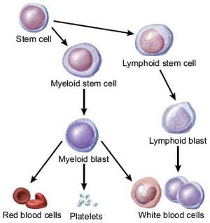
Bone marrow, the soft tissue found inside bones, is the origin point for every cell in the immune system, along with red blood cells and platelets. This all traces back to a single cell type called the hematopoietic stem cell.
Hematopoietic stem cells are capable of both self-renewal, maintaining their own population, and differentiation into any of the specialized blood and immune cell types the body needs. From this starting point, cells branch into two broad lineages. The myeloid lineage produces red blood cells, platelets, and several types of immune cells including neutrophils, macrophages and dendritic cells. The lymphoid lineage produces B cells, T cells and natural killer cells.
B cells complete their maturation directly within the bone marrow, which is in fact where the "B" in B cell comes from, short for bone marrow. T cells follow a different path: they leave the bone marrow as immature precursor cells and travel to the thymus to complete their development, where they undergo the selection process that teaches them to recognize foreign antigens without attacking the body's own tissue.
Because bone marrow is the sole source of these cells, damage to it, whether from certain cancers, chemotherapy or radiation, can severely compromise the entire immune system at once, which is why bone marrow transplants are used to treat conditions including leukemia and certain immune deficiencies, effectively replacing a person's blood and immune cell production system with healthy donor stem cells. The dependence of the entire immune system on this single tissue source is part of why bone marrow health is considered foundational to overall immune function, rather than one component among many operating independently.
-1920x1284.jpg)
Credit: Andrea Piacquadio / Pexels
An allergy is the immune system mounting a defensive response against a substance that poses no actual threat to the body, treating harmless material such as pollen, pet dander or a specific food protein the same way it would treat a genuine pathogen.
The mechanism centers on IgE, the antibody class most associated with allergic reactions. In a person with an allergy, the first exposure to an allergen causes the immune system to produce IgE antibodies specifically shaped to recognize that substance. These IgE antibodies then attach to the surface of mast cells and basophils, two cell types packed with granules containing histamine and other inflammatory chemicals, essentially arming them and priming the body to react on the next exposure.
When the person encounters the same allergen again, the attached IgE antibodies bind it, triggering mast cells and basophils to rapidly release their stored histamine and related chemicals into surrounding tissue. Histamine causes blood vessels to dilate and become more permeable, leading to the swelling, redness and itching typical of an allergic reaction, while also triggering mucus production and, in the airway, constriction of smooth muscle that can cause the wheezing and breathing difficulty associated with allergic asthma.
In severe cases, this same mechanism can occur throughout the body at once, a reaction called anaphylaxis, in which widespread histamine release causes a sudden and dangerous drop in blood pressure along with airway swelling, requiring immediate treatment with epinephrine. Researchers believe this IgE-based system evolved primarily as a defense against parasitic worms, since IgE plays a genuine protective role against those infections; allergic disease is now understood largely as this ancient antiparasitic mechanism misfiring against substances that carry no real threat.
Credit: OpenStax College - Anatomy & Physiology, Connexions Website / Wikimedia Commons (CC BY 3.0)
Regulatory T cells, often abbreviated as Tregs, are a specialized subset of T cells whose job is to suppress immune activity rather than initiate it, acting as a built-in brake on a system that would otherwise be prone to attacking the body's own tissue.
Tregs are identified by a specific internal protein called FOXP3, which acts as a master switch controlling the genes that give these cells their suppressive function. Most Tregs develop in the thymus alongside other T cells, selected specifically for their ability to recognize the body's own proteins, the opposite selection pressure applied to conventional T cells, which are eliminated if they react too strongly to self.
Once active, Tregs use several methods to dial down immune responses. They can directly suppress the activity of other immune cells through cell-to-cell contact, release anti-inflammatory cytokines such as interleukin-10 that counteract pro-inflammatory signaling, and compete with other T cells for access to growth signals needed for those cells to multiply, effectively starving overactive immune responses of the resources they need to expand.
The importance of Tregs becomes clear in their absence. A rare genetic condition in which the FOXP3 gene is nonfunctional, called IPEX syndrome, causes severe, multi-organ autoimmune disease starting in early infancy, since the body has no mechanism to restrain immune cells that react against its own tissue. This condition illustrates that Tregs are not a minor regulatory detail but a core structural requirement for a functioning immune system. Ongoing research into Tregs also extends into cancer treatment and organ transplantation, since boosting or suppressing their activity in targeted ways could help the immune system either attack tumors more aggressively or tolerate a transplanted organ without rejecting it.