
15 things the human body does automatically that most people never think about
From regulating your temperature to cleaning your brain while you sleep, your body runs dozens of critical processes entirely without your knowledge or permission
QAI Publishing / Getty Images
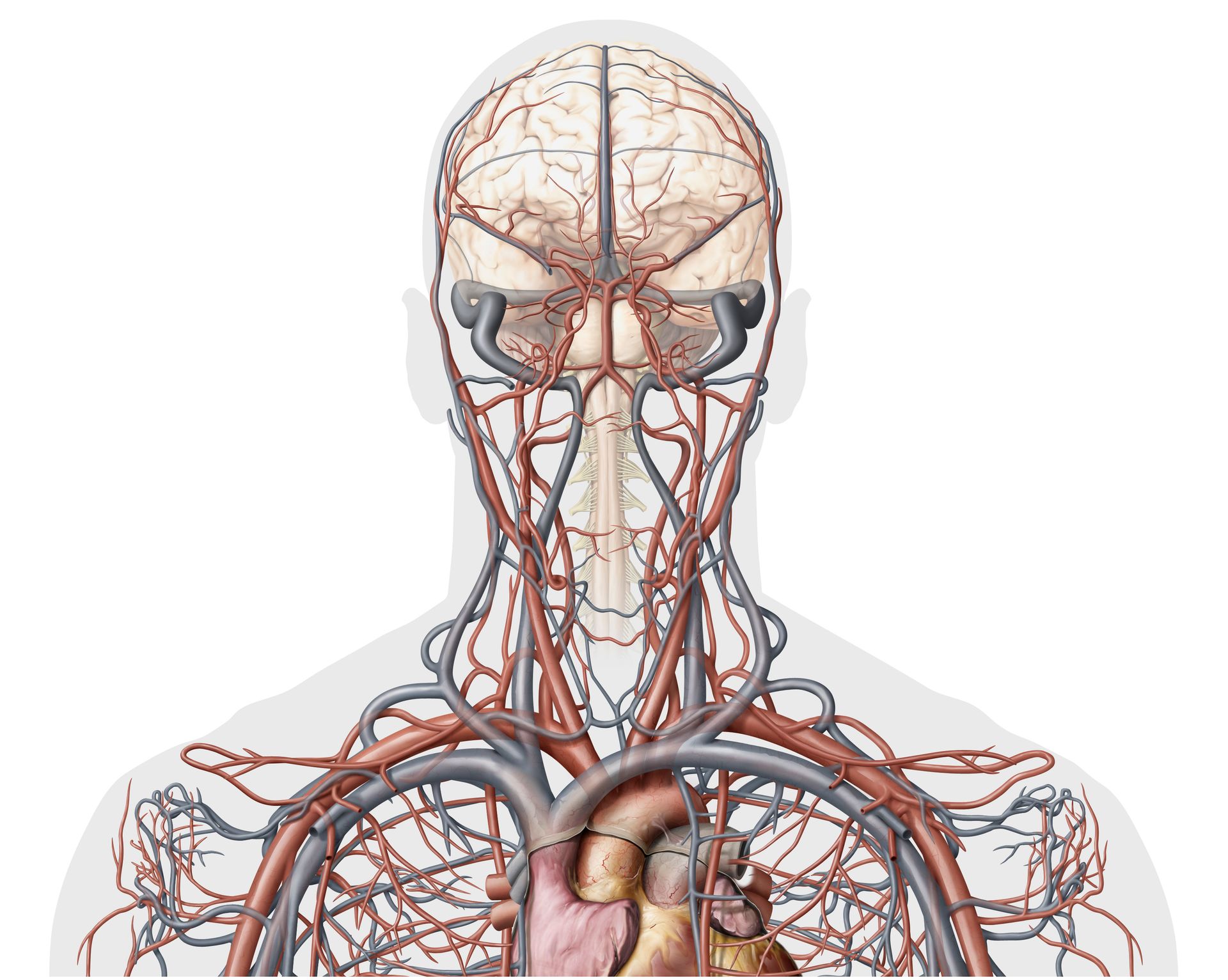
The human body is running roughly 37 trillion cells at any given moment, coordinating systems so intricate and fast that conscious thought would only slow them down. That is why so much of what keeps you alive happens without your permission, your awareness, or even your gratitude.
Most people know the heart beats and the lungs breathe. Those are the famous ones. Below them sits an entire second layer of biological automation — processes that fire, adjust, correct, and reset thousands of times each day without ever surfacing into conscious experience. Your pupils shrink in bright light before your brain has registered the glare. Your blood begins clotting before you have noticed a cut. Your brain washes itself every night while you sleep.
These are not trivial quirks. They are foundational infrastructure. Each one represents millions of years of evolutionary refinement — a solution to a specific survival problem that proved reliable enough to get hard-wired. The gag reflex exists because swallowing the wrong thing could kill you. The diving reflex exists because many mammals, including early humans, regularly found themselves submerged. Goosebumps made functional sense when your ancestors had thick body hair.
Understanding these functions does not just satisfy curiosity. It also offers a useful reminder about the limits of conscious control. When stress disrupts sleep, it interrupts the brain's nightly cleaning process. When anxiety triggers the startle response too frequently, it leaves the nervous system in a state of low-grade readiness that drains energy and impairs judgment. These automatic functions are not separate from your mental and emotional life. They are deeply entangled with it.
This list covers 15 of the body's most consequential automatic processes — the ones that operate entirely beneath the threshold of awareness but shape everything from how clearly you see to how well your blood flows after an injury. Some of them you have probably noticed without ever understanding. Others you have almost certainly never thought about at all.
How your body regulates its own temperature

Hector Vivas / Getty Images
The human body maintains a core temperature of roughly 37 degrees Celsius — 98.6 degrees Fahrenheit — and it does this constantly, through mechanisms that require no conscious instruction. The system is called thermoregulation, and it is managed primarily by a small region of the brain called the hypothalamus, which functions like a thermostat embedded in neural tissue.
When your core temperature rises — from exercise, heat, or a warm environment — the hypothalamus sends signals through the autonomic nervous system to trigger sweating. Sweat glands in the skin release water and salts onto the body's surface. As that water evaporates, it carries heat away from the skin. Blood vessels near the skin's surface also dilate, a process called vasodilation, allowing more blood to flow close to the surface and release heat into the surrounding air. This is why skin turns red during exercise or in hot conditions.
When temperature drops, the system works in reverse. Blood vessels constrict, pulling warm blood away from the extremities and concentrating it around vital organs. This is why fingers and toes go cold first in cold weather. If the temperature continues to fall, the body triggers shivering — rapid, involuntary muscle contractions that generate heat through friction and metabolic activity. Shivering can increase the body's heat production by four or five times the resting rate.
The body also adjusts metabolic rate, releasing hormones like thyroid hormone and adrenaline that ramp up the speed at which cells burn energy, generating more heat as a byproduct. These hormone-driven responses are slower than sweating or shivering but provide sustained adjustment over hours.
The system's precision is worth noting. Core temperature does not need to fluctuate more than a couple of degrees in either direction before serious problems arise. Hypothermia — core temperature below 35 degrees Celsius — begins to impair brain function and organ coordination. Hyperthermia above 40 degrees Celsius can cause heatstroke, a life-threatening condition in which the regulation system itself begins to fail.
The hypothalamus also adjusts its setpoint during illness. A fever is not a malfunction — it is a deliberate recalibration, the hypothalamus raising the target temperature to make the body's environment less hospitable to pathogens. The automatic nature of this process is part of its power: it does not wait for instruction.
The blink reflex and why your eyes never dry out

Chermiti Mohamed / Pexels
Human eyes blink roughly 15 to 20 times per minute on average — a rate that varies with attention, emotion, and fatigue but almost never requires deliberate effort. Over the course of a single waking day, that works out to somewhere between 10,000 and 15,000 blinks. Almost none of them are intentional.
The primary function of blinking is lubrication. Each blink spreads a thin layer of tear film across the surface of the eye, a three-layer structure made up of mucus, water, and oil. The mucus layer, produced by cells in the conjunctiva, helps the tear film adhere to the eye's surface. The watery middle layer, produced by the lacrimal glands, provides hydration and carries oxygen. The oily outer layer, produced by meibomian glands in the eyelids, slows evaporation and keeps the water from dispersing too quickly.
Without blinking, the eye's surface would dry out within seconds in most environments. A dried cornea loses its optical clarity and becomes vulnerable to damage from dust, airborne debris, and friction. The cornea is one of the few tissues in the body with no blood supply — it receives oxygen directly from the air and from the tear film — which makes keeping it moist critical to its function.
Blinking also serves a protective role. The blink reflex — a fast, involuntary closure triggered by sudden movements near the eye, loud noises, or contact with the cornea — can occur within 100 milliseconds, faster than most voluntary movements can be executed. This reflex runs through a circuit involving the trigeminal nerve and the facial nerve, and it operates below the level of conscious processing. By the time you perceive a threat near your eye, your eyelid may already have closed.
Blink rate carries information about cognitive state. It decreases during focused visual tasks — reading, screen use, driving — which is one reason these activities contribute to eye fatigue. Blink rate also decreases during states of high concentration and increases during moments of mental fatigue or distraction.
Blink rate is also linked to dopamine activity in the brain. Changes in blink frequency are observed in several neurological conditions. Parkinson's disease is associated with a reduced blink rate. Schizophrenia tends to show an elevated one. The blink also coordinates with eye movement: the brain suppresses vision momentarily during each blink — a phenomenon called blink suppression — which is why you do not perceive a brief flicker of darkness thousands of times each day.
The gag reflex and what it's actually protecting
cottonbro studio / Pexels
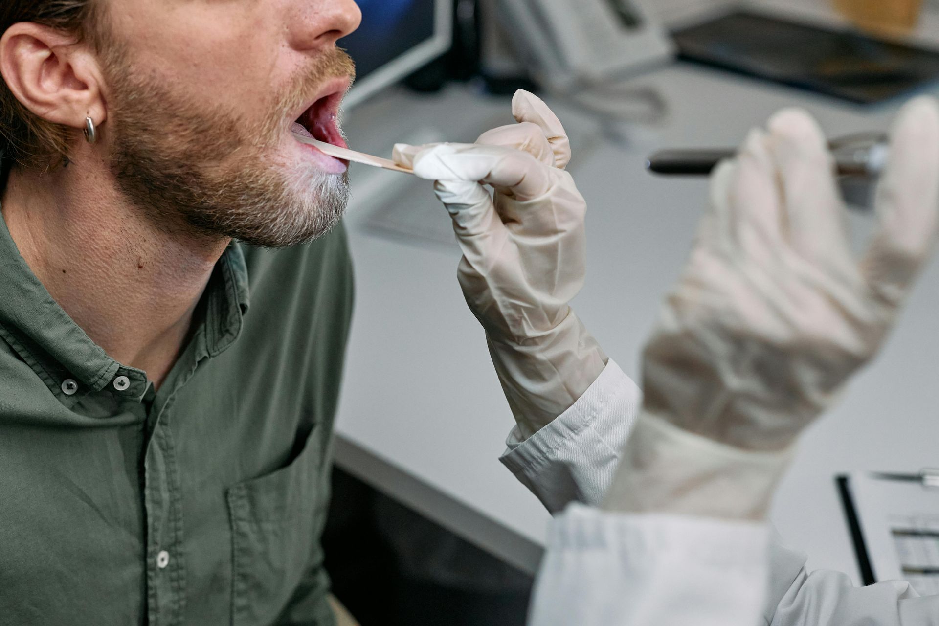
The gag reflex — technically called the pharyngeal reflex — is one of the body's most forceful protective mechanisms. It triggers when something touches the soft palate, the back of the throat, the tonsil area, or the back of the tongue, causing a sudden muscular contraction designed to expel whatever is there. Its purpose is to prevent foreign objects from traveling further into the airway or the esophagus.
The reflex is coordinated by the vagus nerve and the glossopharyngeal nerve — two of the cranial nerves that connect the brainstem directly to the throat and digestive tract. When the appropriate area is stimulated, signals travel almost instantly to the brainstem, which coordinates the muscular response: the soft palate rises, the pharynx contracts, and the back of the tongue retracts. This happens fast enough to intercept objects before they can be aspirated into the lungs.
The sensitivity of the gag reflex varies significantly between individuals — a fact any dentist will confirm. Some people gag easily during routine dental work; others have a suppressed reflex that barely triggers even during throat examinations. This variation reflects differences in nerve sensitivity, anatomy, and psychological conditioning. Anxiety can heighten the reflex's sensitivity, which is why some people gag more easily when they are stressed or tense.
The gag reflex is also affected by what the brain expects. When the brain anticipates stimulation of the throat, the reflex threshold can lower in advance. This is one reason people sometimes gag in response to a sight or smell rather than only direct physical contact — the anticipatory signal primes the mechanism before any physical trigger arrives.
The reflex also differs from the vomiting reflex, though the two are related. Gagging is a local protective response in the throat. Vomiting is a full-body expulsion coordinated by the vomiting center in the medulla oblongata. A gag can trigger vomiting, but gagging does not always result in it. The two involve overlapping but distinct neural circuits.
Age and neurological status affect the reflex's presence. An absent or significantly diminished gag reflex can be a clinical sign of neurological damage — particularly damage to the brainstem or the cranial nerves involved. Clinicians test the reflex by touching the back of the throat with a tongue depressor, using its presence or absence as a rough indicator of brainstem function. The reflex also operates in close coordination with swallowing: once food passes the threshold at the back of the mouth, the rest of the swallow sequence is involuntary, and the gag reflex and swallow reflex trade continuous signals about what is in the throat and what should be done about it.
How your pupils respond to light without any instruction from you
Sergei Starostin / Pexels
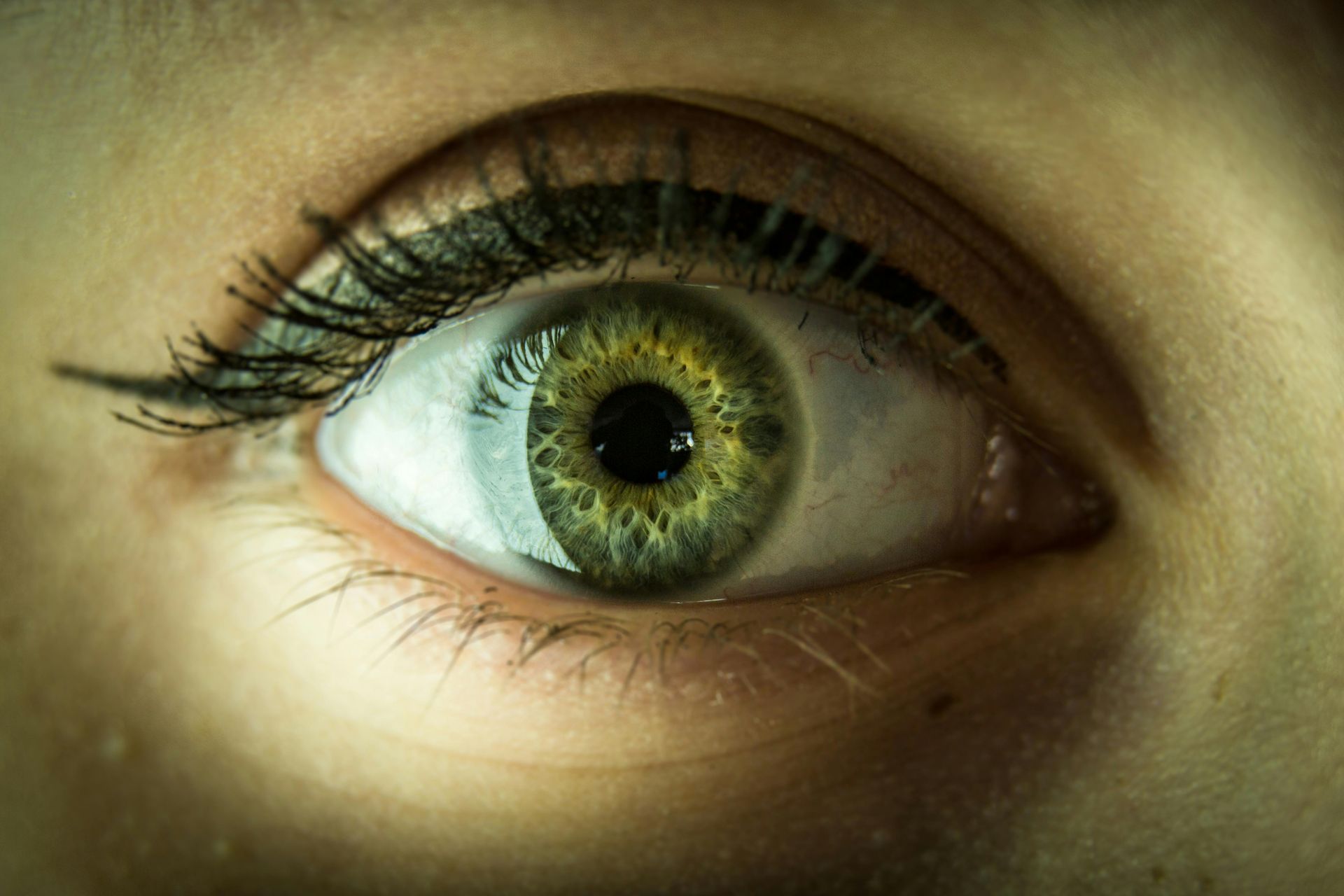
The pupils of the human eye are controlled by two sets of muscles — the sphincter pupillae, which constricts the pupil, and the dilator pupillae, which expands it. Neither muscle is under voluntary control. The entire process is managed by the autonomic nervous system, responding to light, emotional state, and cognitive load in real time.
In bright light, the sphincter pupillae contracts, narrowing the pupil to limit how much light reaches the retina. In low light, the dilator pupillae takes over, widening the pupil to gather more light. The response to a sudden change in light is fast — full constriction in response to a bright flash can occur within a quarter of a second — but sustained adjustment to ambient light levels takes longer, as anyone who has walked from a dark room into sunlight can confirm.
The pupillary light reflex runs through the midbrain rather than the visual cortex, which means it operates independently of conscious vision. A person who is cortically blind — unable to process visual images because of damage to the visual cortex — can still have intact pupillary light reflexes, because the reflex arc bypasses the cortex entirely. This is why doctors use a penlight to check pupil responses in patients with suspected brain injury. Abnormal or asymmetric responses can indicate damage to the midbrain, the brainstem, or the cranial nerves involved.
Pupils also respond to emotional and cognitive states. The sympathetic nervous system — associated with alertness, arousal, and stress — causes pupil dilation independent of light levels. This is why pupils dilate in response to fear, pain, or excitement. They also dilate in response to mental effort: problem-solving, decision-making, and memory retrieval tasks all produce measurable increases in pupil size. Researchers use this response as a non-invasive proxy for cognitive workload in laboratory settings.
Certain drugs strongly affect pupillary response. Opiates cause extreme constriction — pinpoint pupils are a diagnostic sign of opiate overdose. Stimulants like amphetamines cause pronounced dilation. Anticholinergic drugs, which block the nerve signal that triggers constriction, can produce fixed, dilated pupils that no longer respond to light — a medical emergency that requires immediate attention and tells clinicians something specific about which system has been affected.
Peristalsis — the wave your digestive system rides
MART PRODUCTION / Pexels
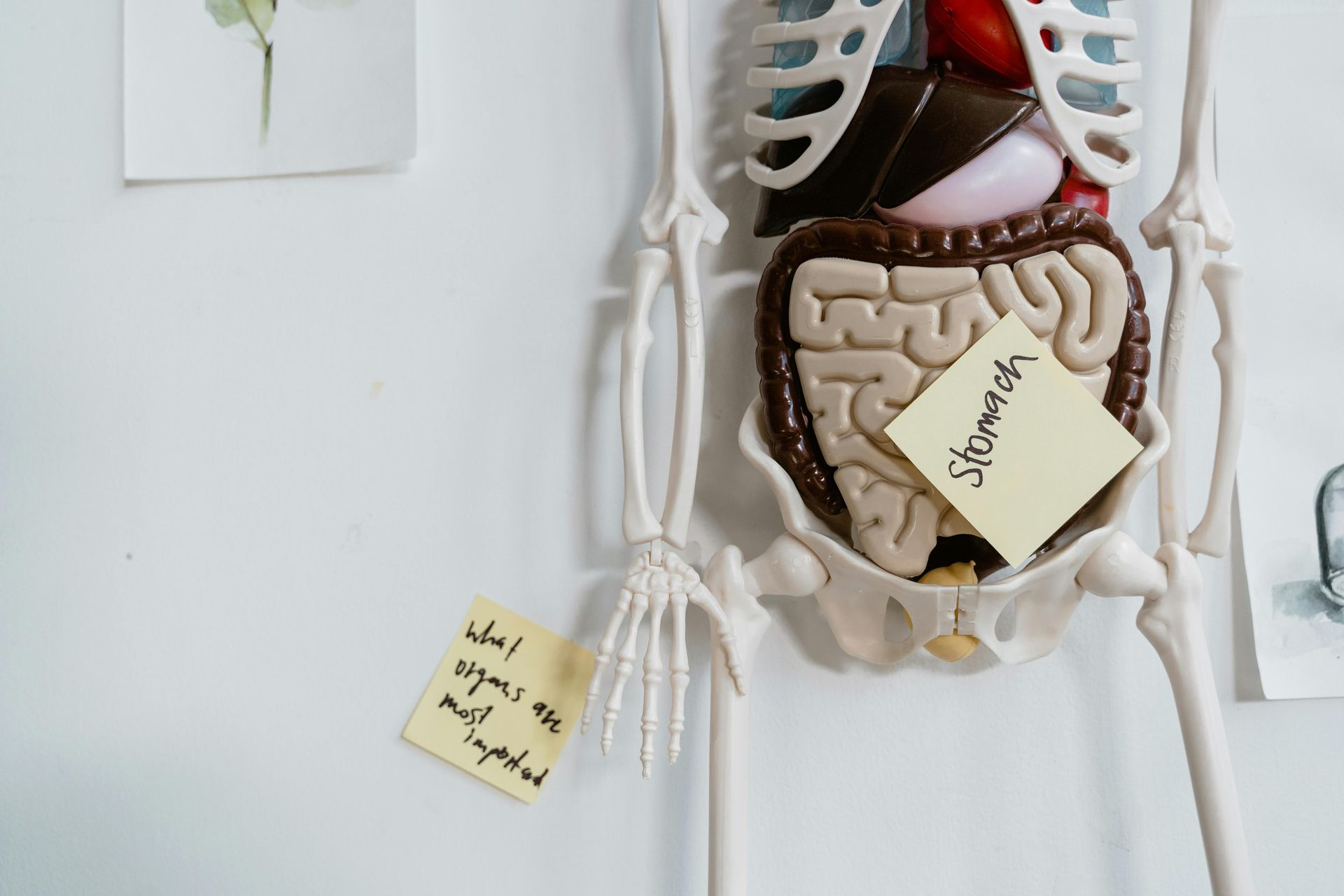
Once you swallow food, you lose all conscious control over where it goes and how fast. The digestive system takes over, moving material through roughly nine meters of tubing using a coordinated, wave-like muscular action called peristalsis.
Peristalsis is produced by two layers of smooth muscle that line the wall of the digestive tract. The circular muscle layer contracts behind a bolus of food, narrowing the tube and pushing the material forward. The longitudinal muscle layer contracts ahead of it, shortening that segment and widening the space. These contractions alternate in sequence, moving the bolus steadily forward. The sequence is controlled not by the brain directly but by the enteric nervous system — a network of roughly 500 million neurons embedded in the walls of the digestive tract, sometimes called the second brain.
The enteric nervous system operates largely independently of the central nervous system. It manages peristalsis, regulates digestive secretions, and coordinates muscular contractions even when its connections to the brain and spinal cord are severed. This autonomy means the gut keeps working during sleep, during stress, and in people with significant spinal cord injuries.
Peristalsis begins in the esophagus. When food is swallowed and the voluntary phase ends, a peristaltic wave moves downward from the top of the esophagus, pushing the bolus toward the stomach. The lower esophageal sphincter — a ring of muscle at the stomach's entrance — relaxes in advance of the wave to allow food through, then closes behind it to prevent stomach acid from flowing back upward.
In the stomach, peristalsis takes a different form. Stronger, more forceful contractions churn the food and mix it with gastric acid and enzymes, breaking it into a semi-liquid paste called chyme. The stomach empties in waves, releasing small amounts of chyme into the small intestine at intervals regulated by the composition of the meal. Fat and protein slow gastric emptying, while simple carbohydrates move through faster.
In the large intestine, peristalsis slows considerably. Mass movements — stronger contractions that occur a few times per day, often triggered by eating — push material toward the rectum. The sensation of urgency before a bowel movement is the result of these mass movements combined with pressure receptors in the rectal wall signaling the brain that material has arrived and evacuation may be warranted. None of this chain of events — from swallow to elimination — requires a single conscious instruction.
The diving reflex — your body's underwater emergency mode

Kindel Media / Pexels
The diving reflex — more precisely called the mammalian diving response — is triggered when the face is submerged in cold water. Within seconds, the body initiates a coordinated set of physiological changes that redistribute oxygen away from the muscles and toward the heart and brain, protecting the organs that need it most.
The first change is bradycardia — a rapid slowing of the heart rate. In regular swimmers, heart rate can drop by 10 to 25 percent within seconds of cold water immersion. In trained divers, the drop can be more pronounced. Submerging the face activates receptors in the nose and around the eyes that send signals through the trigeminal nerve to the brainstem, which responds by increasing parasympathetic outflow to the heart, slowing it down.
Simultaneously, blood vessels in the peripheral parts of the body — the limbs, the skin, and the digestive organs — constrict, a response called peripheral vasoconstriction. This shunts blood toward the core and away from areas where the metabolic demand for oxygen is less critical. The effect is to extend the time the body can survive without breathing by prioritizing oxygen delivery to the brain and cardiac muscle.
The spleen also contracts during the diving response. The spleen stores a reserve of red blood cells, and contracting it releases additional cells into circulation, temporarily increasing the blood's oxygen-carrying capacity. This effect is more pronounced in people who dive regularly — competitive free divers have notably enlarged spleens compared to non-divers, and their spleens contract more forcefully during dives.
The reflex is present in all mammals and is most pronounced in marine mammals like seals and dolphins, which can sustain dives of extraordinary duration. In humans, it is weaker and less consistent, but it is present from birth. It is actually strongest in infants, which is why newborns can be briefly submerged without aspirating water — a property occasionally used in some birthing techniques.
Cold water is essential for triggering the full response. Warm water immersion produces a weaker reflex or none at all. The cold receptors on the face are the primary trigger, which means that covering the face with cold water — even without full-body immersion — is enough to initiate bradycardia in most people. Some free divers use this deliberately, splashing cold water on their face before a dive to pre-activate the response.
Goosebumps and what they were originally for

www.kaboompics.com / Pexels
Goosebumps are produced by tiny muscles at the base of individual hair follicles called arrector pili. When stimulated by the sympathetic nervous system — during cold, fear, or intense emotional experience — these muscles contract, pulling the hair follicle upright. The result is a brief puckering of the skin around each follicle, producing the characteristic bumpy texture named for the appearance of a plucked bird.
In most mammals with dense fur, this reflex serves two practical functions. Raised hair traps a layer of air between the strands, improving the fur's insulating properties and retaining body heat. The same response also makes the animal appear larger, a useful display in confrontations with predators or rivals. In animals with thick coats — cats, bears, porcupines — piloerection is a clearly visible and functionally significant response.
In humans, the reflex has largely lost its practical value. Human body hair is too sparse and fine to trap meaningful amounts of air or create a convincing size display. Goosebumps in response to cold provide no measurable insulation. The mechanism persists as an evolutionary remnant — a reflex inherited from heavily-furred ancestors — that the body runs without any functional benefit in its current context.
The emotional trigger for goosebumps is more interesting. Piloerection in response to music, emotional storytelling, awe, or nostalgia — sometimes called chills, or the German term frisson — activates through a partially overlapping but distinct neural circuit from the cold-triggered version. Both involve the sympathetic nervous system, but frisson appears to be connected to the brain's reward pathways. People who experience music-induced chills show measurable dopamine release in the striatum during those moments.
Not everyone experiences frisson. It appears to be more common in people who score high on the personality trait of openness to experience, and the capacity for it is at least partly heritable. Some people experience it frequently; others almost never, even when exposed to music or emotional content they find deeply affecting.
The temperature-triggered version of piloerection operates through a different pathway — primarily through cold receptors in the skin sending signals to the spinal cord, which activates the sympathetic chain. It does not require conscious perception of cold. The reflex can be triggered during anesthesia, as long as the skin temperature drops sufficiently to activate the relevant receptors. The muscles are ready and waiting, regardless of what the conscious mind is doing.
Blood clotting — the body's emergency sealing system

MART PRODUCTION / Pexels
The moment a blood vessel is damaged, the body begins a complex, multi-stage repair process designed to stop bleeding without permanently blocking the vessel. The process, called hemostasis, runs through three overlapping phases that together produce a clot, seal the breach, and then — in most cases — dissolve the clot once the vessel has healed.
The first phase begins within seconds. Damaged blood vessels release chemical signals that cause the vessel to contract, reducing blood flow to the injured area. Simultaneously, specialized cell fragments called platelets, which circulate continuously in the blood, detect the damage. Platelets normally slide past the smooth inner lining of blood vessels without sticking, but when the vessel wall is torn, proteins in the underlying tissue — particularly collagen — are exposed. Platelets bind to collagen, become activated, change shape, and release chemical signals that recruit more platelets to the site. Within minutes, a soft plug of platelets accumulates at the breach.
This platelet plug is enough to seal minor cuts in small vessels. For larger injuries, the body needs a more durable structure, which is where coagulation comes in. Coagulation is a cascade: a series of proteins in the blood called clotting factors activate each other in a sequence, each step amplifying the signal so that a small initial trigger leads rapidly to a large, coordinated response. The cascade's endpoint is the production of thrombin, an enzyme that converts a soluble blood protein called fibrinogen into fibrin. Fibrin strands weave together into a mesh that reinforces the platelet plug, trapping red blood cells and forming a firm, stable clot.
The body also runs a parallel system designed to prevent clotting from spreading beyond the injury site. Natural anticoagulants in the blood — proteins like antithrombin and protein C — limit the activity of clotting factors to the immediate area of damage. This balancing act is critical. Too little clotting causes hemorrhage; too much causes thrombosis, the formation of clots inside intact vessels, which can block blood flow to the heart, lungs, or brain.
Once the vessel heals, a process called fibrinolysis breaks down the clot. The enzyme plasmin gradually dissolves the fibrin mesh, clearing the vessel and restoring normal blood flow. The entire system operates without conscious input at any stage — from the first chemical signal at the moment of injury to the final dissolution of the clot days or weeks later.
How your body knows when to breathe
Aakash Dhage / Unsplash
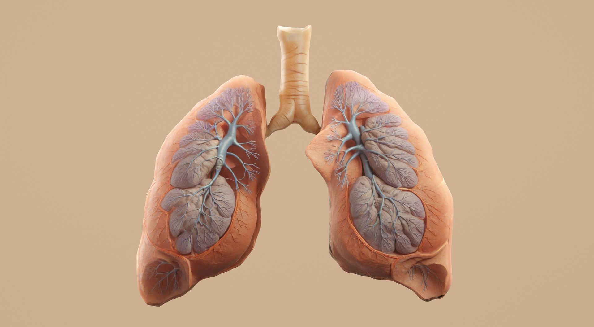
Breathing is one of the few automatic functions that can also be consciously controlled — you can hold your breath, slow it down, or deliberately speed it up. But the automatic system that manages breathing under normal conditions does not rely on conscious command, and it overrides voluntary control when circumstances require it.
The primary driver of automatic breathing is not oxygen level but carbon dioxide. Specialized chemoreceptors in the medulla oblongata — a part of the brainstem — continuously monitor the concentration of carbon dioxide in cerebrospinal fluid and in the blood. When carbon dioxide rises above a certain threshold, these receptors send signals to the respiratory centers in the brainstem, which in turn signal the diaphragm and intercostal muscles to contract. You inhale, bringing in fresh air, and then exhale, expelling carbon dioxide. As CO2 levels fall back toward normal, the signaling eases, and breathing slows or pauses briefly until CO2 builds again.
Peripheral chemoreceptors — located in the carotid bodies near the carotid arteries and in the aortic bodies near the aorta — also monitor oxygen levels. These receptors become active when blood oxygen falls significantly, triggering increased respiratory drive. But under normal conditions, the CO2 system is more sensitive and more tightly regulated than the oxygen system. The body tolerates relatively large swings in blood oxygen before the peripheral chemoreceptors trigger a strong response, whereas even small rises in CO2 produce rapid adjustments in breathing rate and depth.
This is why hyperventilating — breathing rapidly and deeply for an extended period — makes you feel dizzy and can cause loss of consciousness. Hyperventilation does not raise oxygen levels significantly, since blood is already nearly saturated under normal conditions. But it does lower CO2 rapidly. With CO2 suppressed, the brainstem's drive to breathe weakens, and in extreme cases the urge to breathe disappears temporarily.
The same mechanism explains why competitive breath-hold divers sometimes lose consciousness underwater. They hyperventilate before a dive to suppress the CO2-driven urge to breathe, extending the time they can stay submerged. But they may lose consciousness from oxygen deprivation before CO2 rises enough to drive them to the surface — a condition known as shallow-water blackout.
The respiratory control system also adjusts for altitude, exercise, sleep, and acid-base balance. During exercise, CO2 production rises sharply, and breathing increases in proportion. During sleep, the system's sensitivity changes, which is why breathing becomes slower and shallower, and why sleep apnea — an interruption of breathing during sleep — is a distinct clinical condition requiring its own treatment.
The startle response

Andrea Piacquadio / Pexels
The startle response is one of the fastest reflexes in the human nervous system. An unexpected loud sound, a sudden movement in peripheral vision, or an abrupt touch triggers a cascade that begins in the brainstem and produces a full-body response in less than 150 milliseconds — faster than a conscious decision about any action could possibly be made.
The response has a characteristic physical signature. The eyes close, the facial muscles tighten, the shoulders rise, the arms flex and pull inward, the knees bend slightly, and the head ducks forward. This posture is thought to protect the body's most vulnerable areas — the eyes, the throat, and the major blood vessels at the neck — while orienting the body toward a possible threat.
The neural pathway runs through the reticular formation in the brainstem, bypassing the cortex. This is why the startle response is extremely difficult to suppress voluntarily, even when you know a startling stimulus is coming. The signal reaches the brainstem and activates the response before cortical processing — which includes the recognition that the noise is, for instance, a car backfiring rather than a weapon — can intervene. The cortex can reduce the response over time through habituation, which is why repeated exposure to the same stimulus produces a smaller and smaller reaction, but on first exposure, the reflex fires almost unconditionally.
The amygdala, the brain region most associated with threat processing, is not required for the initial startle response — that runs through the brainstem alone. But the amygdala does influence the intensity and duration of the response. People with heightened amygdala activity — as seen in post-traumatic stress disorder — show exaggerated startle responses that are harder to habituate. This exaggerated startle is one of the diagnostic criteria for PTSD, reflecting a nervous system that has recalibrated its threat sensitivity upward.
The startle response is present at birth. Newborns show a version of it — called the Moro reflex — in which the arms extend outward and then pull inward in response to a sudden loud sound or the sensation of falling. The Moro reflex fades within the first few months of life as the developing cortex gains more influence over brainstem activity, but the underlying neural architecture that supports the adult startle response is present from the earliest weeks of life.
Acoustic startle — triggered by sound — is the most studied form, and the ease with which it can be measured has made it a useful tool in neuroscience research. By measuring the magnitude and habituation rate of the startle response, researchers can draw inferences about the functioning of the brainstem, the amygdala, and various neurotransmitter systems, including those affected by psychiatric medications and conditions.
The circadian clock that runs every cell in your body
-1920x1280.jpg)
Anastasiya Vragova / Pexels
Every cell in the human body contains a molecular clock — a set of proteins that cycle through activation and suppression on a roughly 24-hour schedule. This cellular timekeeping system, called the circadian clock, coordinates nearly every biological process in the body to a specific time of day: when to release certain hormones, when to consolidate memories during sleep, when to raise core temperature, when to trigger peak alertness, and when to initiate the cellular repair processes that occur during rest.
The master clock sits in a small cluster of neurons in the hypothalamus called the suprachiasmatic nucleus, or SCN. The SCN receives input from specialized light-sensitive cells in the retina — cells that respond primarily to blue-wavelength light and that are distinct from the rods and cones used for vision. This light input resets the clock each day, synchronizing the body's internal rhythm to the actual solar cycle.
When the SCN detects light at the expected time — morning — it suppresses melatonin production by the pineal gland and triggers a gradual increase in cortisol, raising alertness. Body temperature begins to rise. Blood pressure increases. The digestive system activates. Insulin sensitivity improves in anticipation of eating. In the evening, as light fades, melatonin production begins, core temperature drops, and the body transitions toward sleep.
The circadian system coordinates peripheral clocks in every organ — the liver, the heart, the lungs, the immune system — and disrupting the synchronization between these clocks and the master SCN clock has measurable health consequences. Shift workers, frequent long-haul travelers, and people who consistently stay up late while exposed to artificial light show elevated rates of metabolic dysfunction, cardiovascular disease, and impaired immune response. This is not just about feeling tired — it reflects a genuine desynchronization of biological processes that evolved to operate in coordination.
The body can run its clock independently of light input for some time. People in constant darkness will maintain a roughly 24-hour rhythm, though it drifts slightly. This reflects the fact that the intrinsic period of the human clock is close to but not exactly 24 hours. Light input from the environment corrects this drift each day, keeping the body synchronized with the external world — a recalibration that happens every morning without any conscious participation.
The tears you never notice

Sean White / Getty Images
Most people think of tears as something produced during crying. The eye produces tears continuously — every second of every waking hour, and at a reduced rate during sleep. These are called basal tears, and they are a critical component of eye health, not an emotional response.
Basal tears are produced by the lacrimal glands, located above and to the outer side of each eye, as well as by smaller accessory glands distributed through the conjunctiva. The glands release a steady, low-volume flow of fluid that the eyelids spread across the eye's surface with each blink. Most of this fluid drains through small openings at the inner corner of the eye — the puncta — into the nasolacrimal duct, which carries it into the nasal cavity. This is why crying produces a runny nose: the tear volume exceeds the drainage capacity, and fluid backs up and spills down the cheek while also draining into the nose.
Basal tears serve multiple functions simultaneously. They maintain the optical clarity of the cornea by keeping it smooth and uniformly hydrated. They carry oxygen and nutrients to the corneal epithelium, which lacks blood vessels. They contain lysozyme, an enzyme that breaks down bacterial cell walls and provides continuous low-level antimicrobial protection. They flush out small particles, dust, and irritants before those materials can abrade the corneal surface.
The composition of basal tears differs from emotional tears. Basal tears are primarily water, salts, proteins, and oils. Emotional tears contain higher concentrations of certain hormones and proteins, including prolactin, adrenocorticotropic hormone, and leucine enkephalin — a natural pain-reducing compound. Some researchers have proposed that emotional crying may serve a partial excretory function, releasing stress hormones through the tear ducts. The evidence is suggestive but not conclusive.
Reflex tears — produced in response to an irritant like smoke, onion vapor, or a foreign object — are the third category. These are produced rapidly and in large volume specifically to dilute and flush the irritant from the eye's surface. The trigger runs through the trigeminal nerve: sensory signals from the cornea travel to the brainstem, which signals the lacrimal glands to increase output sharply.
The automatic production of basal tears is so continuous and unremarkable that most people are entirely unaware of it. Yet dry eye disease — in which basal tear production is insufficient or tear film quality is degraded — produces significant daily discomfort and, in severe or untreated cases, corneal damage. The absence of something the body was doing invisibly becomes noticeable only when it stops working.
Proprioception — the sense that tells you where your body is

Yan Krukau / Pexels
Close your eyes, hold out your arm, and touch your index finger to your nose. You can do this easily without visual feedback, because your brain receives a continuous stream of information about the position, movement, and tension of every joint and muscle in your body — a sense called proprioception.
Proprioception is delivered by specialized sensory receptors distributed throughout muscles, tendons, and joint capsules. Muscle spindles — sensory fibers embedded in muscle tissue — detect changes in muscle length and the speed at which that length changes. Golgi tendon organs, located where muscles meet tendons, monitor tension. Mechanoreceptors in joint capsules provide information about joint angle and loading. Signals from all these receptors travel continuously to the spinal cord and brain, where they are integrated into a real-time model of the body's position and movement.
Much of this processing happens below the level of conscious awareness. The cerebellum — a structure at the back of the brainstem — receives proprioceptive information and uses it to fine-tune motor commands before they reach the muscles. This is what allows fluid, coordinated movement without constant deliberate effort. When you catch a ball, shift your weight on uneven ground, or adjust your grip on an object that starts to slip, the cerebellum and spinal cord handle most of the corrective action before your conscious mind has fully registered that a correction was needed.
Proprioception interacts closely with the vestibular system — the balance organs in the inner ear — and with vision. Together, these three inputs give the brain the information it needs to maintain balance and spatial orientation. When they conflict — as they do during seasickness, when the inner ear detects motion but the visual environment suggests stillness — the result can be nausea and disorientation.
Proprioceptive function declines with age. Older adults show measurable decreases in joint position sense and balance, which contribute to the elevated fall risk associated with aging. Peripheral neuropathy — damage to the peripheral nerves caused by diabetes, chronic alcohol use, or certain medications — also impairs proprioceptive input, producing the wide-based, unsteady gait characteristic of people who have lost reliable feedback from their feet and lower legs.
The system can also be deceived. The rubber hand illusion — in which a person watches a rubber hand being stroked while their real hand, hidden from view, is stroked in synchrony — causes people to feel that the rubber hand is their own. The illusion demonstrates how powerfully the brain integrates visual and tactile signals with proprioceptive information, and how readily that integration can be manipulated by conflicting sensory input.
The fever response — a deliberate act, not a malfunction
Credit: www.kaboompics.com
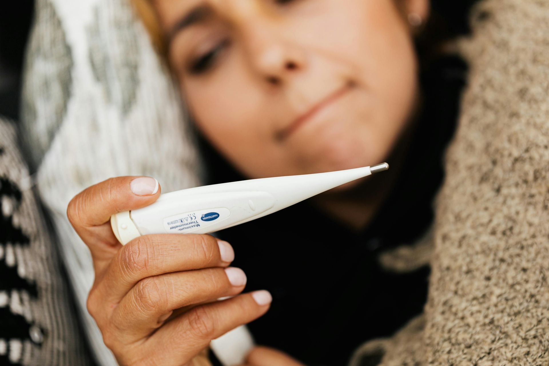
Fever is the body's deliberate elevation of core temperature in response to infection, inflammation, or injury. It is not a malfunction or a side effect of illness. It is an active, energy-intensive defensive strategy, coordinated by the immune system and the brain.
When the body detects pathogens or tissue damage, immune cells release signaling molecules called pyrogens — including interleukins and tumor necrosis factor. These molecules travel to the hypothalamus, where they trigger the production of prostaglandin E2, a lipid compound that acts directly on the temperature-regulating centers of the hypothalamus. The result is a raising of the temperature setpoint: the body's thermostat is turned up.
With the setpoint elevated, the body uses its thermoregulatory mechanisms to reach the new target. It constricts peripheral blood vessels, reducing heat loss at the skin. It triggers shivering to generate heat through muscle activity. Core temperature rises toward the new setpoint and stabilizes there. This is the hot, flushed phase of fever that follows the earlier chills — the chill phase is the body working to reach the elevated target, and the flush phase is the body maintaining it.
The elevated temperature has several effects on the immune response. Many bacteria and viruses replicate less efficiently at higher temperatures. They have evolved to thrive at normal human body temperature, and a sustained fever of 38.5 or 39 degrees Celsius disrupts their metabolic processes. Fever also accelerates aspects of the immune response itself. Neutrophils and macrophages — white blood cells that engulf pathogens — move more quickly and kill more efficiently at elevated temperatures. Natural killer cells become more active. The production and release of certain immune proteins accelerates.
Antipyretic drugs — fever reducers like ibuprofen and acetaminophen — work by blocking the production of prostaglandin E2. This removes the hypothalamus's instruction to maintain the elevated setpoint, and the body then uses its cooling mechanisms — sweating and vasodilation — to bring temperature back down.
Fever becomes medically dangerous at very high temperatures — generally above 40 to 41 degrees Celsius — at which point proteins in cells can begin to denature and normal cellular function is threatened. The brain is particularly vulnerable to extreme heat. The body has protective mechanisms that limit how high a fever can climb, but these can be overwhelmed in severe infections or certain toxic conditions, making very high fevers a genuine medical emergency.
How the brain cleans itself while you sleep

Ron Lach / Getty Images
The brain produces metabolic waste during ordinary activity — proteins, cellular debris, and other byproducts of neurotransmission and energy use. During waking hours, this waste accumulates. The brain lacks a conventional lymphatic system of the kind that clears waste from other organs, so it uses a different system — one that operates primarily during sleep.
This system is called the glymphatic system, a term coined by neuroscientist Maiken Nedergaard and colleagues following their description of its function in 2013. The system works by pumping cerebrospinal fluid through channels surrounding blood vessels in the brain tissue, flushing out accumulated waste products and carrying them toward the liver for disposal.
During sleep, support cells in the brain called astrocytes shrink slightly, expanding the spaces between cells by roughly 60 percent compared to the waking state. This expansion dramatically increases the flow of cerebrospinal fluid through brain tissue. The fluid moves through channels called perivascular spaces, surrounding the arteries that penetrate the brain, and exits through venous channels that connect to the lymphatic system in the neck.
One of the waste products the glymphatic system clears is amyloid beta — a protein that, when it accumulates in the brain, forms the plaques associated with Alzheimer's disease. Sleep deprivation consistently raises amyloid beta levels in cerebrospinal fluid, and over time, chronic poor sleep is associated with higher rates of amyloid plaque formation. The connection between sleep disruption and neurodegenerative disease is an active area of research, and the glymphatic system is central to it.
The system is most active during slow-wave sleep — the deepest stage — and is significantly less active during REM sleep and barely active while awake. This means that total sleep duration, and particularly the duration of deep sleep, matters for how thoroughly the brain's waste clearance is completed each night.
The glymphatic system also clears tau protein, another molecule implicated in Alzheimer's disease and related conditions. Disruptions to glymphatic function have been linked not only to neurodegeneration but also to the aftermath of traumatic brain injury, where waste clearance may already be impaired.
For most of human history, the brain had no known lymphatic drainage equivalent. The discovery of the glymphatic system changed how neuroscientists understand the relationship between sleep and brain health, and it provides a partial explanation for why a night of poor sleep leaves the mind feeling slow and clouded in a way that is, at least in part, chemical — the debris of the previous day's thinking, insufficiently cleared.