cottonbro studio / Pexels
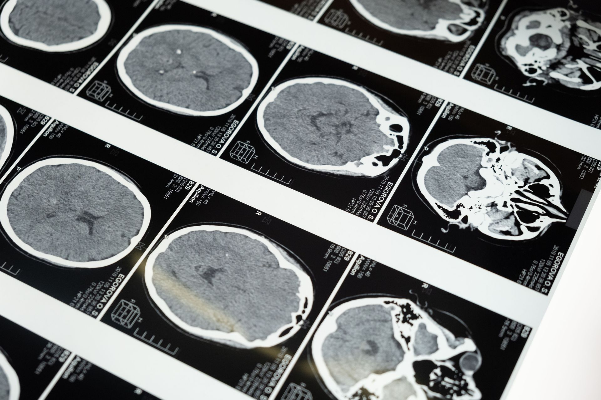
For most of human history, the brain was the organ that could not study itself. The methods available for investigating brain function were limited to the consequences of brain damage — what a patient lost when a specific region was destroyed — and to the post-mortem examination of tissue that had already stopped working. These methods produced genuine knowledge: the localization of language to Broca's and Wernicke's areas in the 19th century, the mapping of the motor and sensory cortices, the broad outlines of neural anatomy. But they were the knowledge of a structure studied from outside, the way you might understand a factory by watching what breaks down when individual machines are removed.
The revolution of the last three decades came from tools that allowed observation of the living, working brain: functional magnetic resonance imaging (fMRI), which detects the blood-flow changes that accompany neural activity; electroencephalography at resolutions that could track the timing of neural events with millisecond precision; optogenetics, which allowed researchers to turn specific neurons on and off with light; and the sequencing and genomic analysis of individual brain cells at scale. These tools did not merely refine the existing picture of the brain. They overturned large parts of it.
The specific overturnings are what this list covers. The brain was thought to be largely fixed in structure after early childhood — it is not. It was thought to generate no new neurons in adulthood — it does, in at least one region. It was thought to be a relatively passive processor of sensory input — it is more accurately described as a prediction engine that continuously generates expectations and updates them against incoming data. The unconscious was thought to be a Freudian domain of repressed wishes — it is better understood as a vast, rapid, mostly efficient processing system that handles far more of cognition than consciousness does.
Each entry in this list is based on peer-reviewed research and represents a finding that was either not known or was thought to be the opposite of true a generation ago. Where findings remain contested or are at the frontier of the evidence base, that is noted. The goal is the specific satisfaction of understanding something as extraordinary as the organ in your skull, with the accuracy that the research now supports.
1 / 25

Caleb Oquendo / Pexels
The dominant model of brain function for most of the 20th century was the input-output model: sensory organs detect the external world, transmit signals to the brain, and the brain processes these signals to produce perception and behavior. The current understanding, developed through the work of Karl Friston, Andy Clark, Jakob Hohwy, and others in the framework called predictive processing or active inference, is almost the reverse: the brain primarily generates predictions about what it expects to encounter and updates those predictions when the incoming sensory data doesn't match.
The mechanism: the brain maintains a hierarchical model of the world, from the most abstract (general principles about how the world works) to the most specific (what the left edge of this desk feels like under my hand right now). This model generates predictions at every level, and what the brain primarily processes is the prediction error — the discrepancy between what was expected and what actually arrived. When prediction errors are large, attention is directed to the discrepancy, the model is updated, and new predictions are generated. When prediction errors are small, the existing model is confirmed and behavior can proceed efficiently.
This model explains a large number of phenomena that the input-output model could not: why perception is so strongly influenced by expectation, why the brain fills in sensory gaps rather than leaving them blank, why chronic pain can persist after tissue damage has healed (the pain prediction has become self-sustaining), and why illusions work (the brain's model is wrong in a specific way that sensory input cannot easily correct). The predictive processing framework is not universally accepted — it is a live theoretical debate — but it is the most influential current framework for understanding how the brain generates experience.
2 / 25

Los Muertos Crew / Pexels
For most of the 20th century, neuroscience held that the brain's structure was largely fixed after a critical period in early childhood — that the neural connections established in the first years of life were essentially permanent and that adult learning worked within this fixed architecture rather than changing it. This view has been comprehensively overturned.
The modern understanding of neuroplasticity — the brain's ability to reorganize its structure and function in response to experience, learning, and injury throughout life — is one of the most practically significant findings in neuroscience. Experience changes the brain. Learning new skills produces measurable changes in the size and connectivity of relevant brain regions. Sustained mental practices — meditation, musical training, taxi driving, second language acquisition — produce structural brain changes visible on MRI. Injury to one region can result in the reorganization of other regions to partially compensate for the lost function.
The most dramatic demonstrations of adult plasticity come from studies of London taxi drivers (Maguire et al., 2000), whose posterior hippocampus — the region associated with spatial navigation — was measurably larger than that of matched controls, with the enlargement correlating with years of experience. Similar structural changes have been found in musicians (enlarged motor and auditory regions corresponding to their instrument), meditators (thickened prefrontal cortex), and people learning to juggle.
The practical implication is that the brain is not a fixed resource whose quality is determined at birth and declines with age. It is a dynamic system that is continuously shaped by what it does — a finding that has changed both how stroke rehabilitation is approached (intensive practice can recruit new circuits to compensate for damaged ones) and how aging and cognitive decline are understood.
3 / 25

Yan Krukau / Pexels
The generation of new neurons — neurogenesis — was thought for most of the 20th century to be restricted to fetal and early childhood development. The adult brain, it was assumed, worked with a fixed complement of neurons that could not be replaced when lost. This assumption began to be challenged in the 1990s, and by the early 2000s, adult neurogenesis in the hippocampus — specifically in the dentate gyrus region of the hippocampus — was established in rodents and later in humans.
The specific finding: new neurons are generated in the hippocampal dentate gyrus in adult humans, and these new neurons are incorporated into existing circuits and contribute to certain types of learning and memory, particularly the formation of new episodic memories and the ability to distinguish between similar experiences (pattern separation). The rate of neurogenesis appears to be regulated by experience: exercise, environmental enrichment, and learning increase neurogenesis; stress, social isolation, and alcohol decrease it.
Whether adult neurogenesis occurs in other brain regions remains contested — some research has proposed neurogenesis in the olfactory bulb and prefrontal cortex, but the evidence is less clear than for the hippocampus. A 2018 study in Nature by Sorrells and colleagues challenged even hippocampal neurogenesis in adult humans, finding no evidence of new neurons in adult hippocampal tissue. The controversy has not been resolved, and the question of the extent and functional significance of adult human neurogenesis is actively debated.
What is not contested is that the adult brain is far more dynamic than the no-neurogenesis model assumed, and that the cellular turnover, synaptic remodeling, and circuit reorganization that occur throughout adult life represent forms of structural change that were not recognized a generation ago.
4 / 25
Mart Production / Pexels
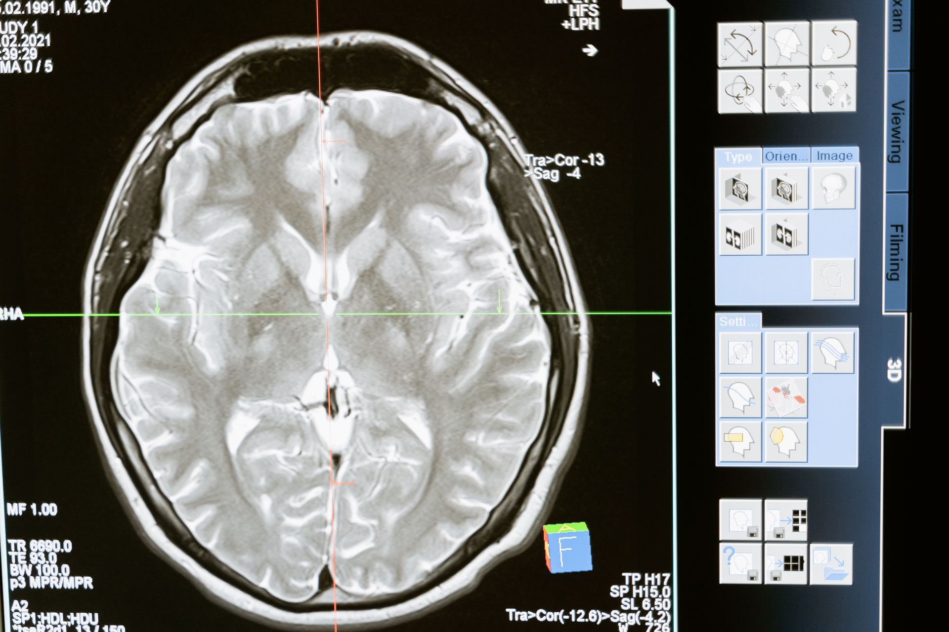
Before the era of neuroimaging, the brain was studied primarily in terms of what it did during specific tasks — what regions activated when a person read, calculated, moved their hand, or processed a face. The assumption was that the brain was essentially inactive between tasks. The discovery of the default mode network (DMN) overturned this assumption and revealed a large, coordinated network of brain regions that is specifically active when the brain is not engaged in a specific external task — during rest, daydreaming, mind-wandering, and self-referential thought.
The DMN — which includes the medial prefrontal cortex, the posterior cingulate cortex, and the angular gyrus, among other regions — was first formally described by Marcus Raichle and colleagues in 2001, based on the observation that these regions consistently deactivated during goal-directed tasks and activated during rest. Subsequent research revealed that the DMN is not idle during rest but engaged in a specific and important set of processes: autobiographical memory retrieval, mental simulation and future planning, perspective-taking, social cognition, and the construction of narrative self-identity.
The DMN's disruption has since been associated with several neurological and psychiatric conditions. Depression is associated with hyperactivity of the DMN — the rumination characteristic of depression appears to involve excessive default mode activity directed at the self. Mind-wandering during a task involves the DMN overriding the task-relevant network. And the disintegration of DMN coherence is one of the earliest signatures of Alzheimer's disease detectable by neuroimaging.
The discovery of the DMN fundamentally changed the model of the resting brain from an inactive state to an active internal processing mode with specific functions, and it changed how neuroimaging studies were designed and interpreted.
5 / 25

Polina / Pexels
The function of sleep — why animals, including humans, spend approximately a third of their lives unconscious and vulnerable — was one of the longest-standing mysteries in biology. The traditional understanding was that sleep was primarily restorative: the body and brain recovered from the demands of wakefulness. The modern understanding identifies a much more specific function: sleep is when the brain consolidates memories, moving information from short-term hippocampal storage to long-term cortical storage, and when it performs a literal cleaning of metabolic waste products.
The memory consolidation finding — developed through the work of Matthew Walker at the University of California, Berkeley, Robert Stickgold at Harvard, and many others — demonstrates that sleep is an active process of memory processing. During slow-wave sleep, the hippocampus replays experiences encoded during wakefulness, and the repeated replay drives the transfer of information to the neocortex for long-term storage. During REM sleep, the brain processes emotional memories and integrates new information with existing knowledge, producing the dream content that reflects the day's experiences recombined with older memories.
The glymphatic system — discovered by Maiken Nedergaard at the University of Rochester in 2013 — revealed a second critical sleep function: the clearance of metabolic waste products from the brain, including amyloid-beta and tau proteins that accumulate in the brain during wakefulness and are associated with Alzheimer's disease. The glymphatic system operates primarily during deep sleep, and its activity represents the brain's own cleaning mechanism — one that is disrupted by sleep deprivation.
The practical implication is that sleep deprivation is not merely fatigue — it impairs memory formation, disrupts emotional regulation, and prevents the clearance of neurotoxic waste products.
6 / 25

Katrin Bolovtsova / Pexels
Mirror neurons — neurons that fire both when an animal performs an action and when it observes the same action performed by another — were discovered in the early 1990s by Giacomo Rizzolatti and colleagues at the University of Parma while studying the motor cortex of macaque monkeys. The discovery was accidental: a neuron was recorded from a monkey that fired when the monkey reached for food, and was found to also fire when a researcher reached for food in the monkey's presence.
The human mirror neuron system, inferred from neuroimaging rather than direct recording (single-neuron recording in healthy humans is not routinely possible), has been proposed as the neural basis of imitation, empathy, language, and social understanding. The most ambitious proposal — Vilayanur Ramachandran's description of mirror neurons as "the neurons that shaped civilization" — generated enormous popular interest and significant scientific controversy.
The controversy is real and important. The evidence for human mirror neurons is indirect; the leap from motor imitation to empathy and social cognition involves many untested assumptions; and the claimed deficits of the mirror neuron system in autism (the "broken mirror" hypothesis) have not been consistently supported by subsequent research. The mirror neuron system is genuine and interesting; the specific functions attributed to it by popular accounts are significantly overstated.
What the mirror neuron research has contributed is the understanding that the motor system is not purely an output system but is involved in the perception and understanding of others' actions — that watching someone act activates the circuits that would produce the same action in the observer, which has genuine implications for social cognition and for understanding imitation learning.
7 / 25
Mart Production / Pexels
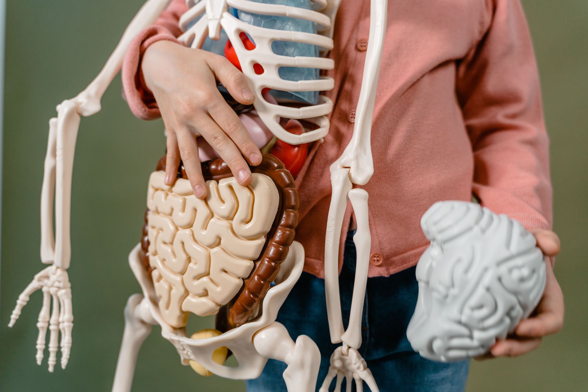
The bidirectional communication system connecting the gut and the brain — the gut-brain axis — is one of the most significant recent expansions of neuroscience, establishing that the enteric nervous system (the approximately 500 million neurons lining the gastrointestinal tract) and the gut microbiome (the trillions of microorganisms in the gut) communicate with the brain in ways that influence mood, cognition, and behavior, and that have been linked to depression, anxiety, autism spectrum disorder, and Parkinson's disease.
The vagus nerve — the primary pathway of the parasympathetic nervous system — carries approximately 80 to 90% of its signals upward, from the gut to the brain, rather than downward. This means the gut is a primary source of information to the brain rather than primarily receiving instructions from it. The gut microbiome produces neurotransmitter precursors (approximately 95% of the body's serotonin is produced in the gut), metabolites that cross the blood-brain barrier and influence neuronal function, and immune signals that modulate neuroinflammation.
The finding that the gut microbiome influences brain function and behavior has been demonstrated in animal studies with particular clarity: germ-free mice (raised without any gut microbiome) show exaggerated stress responses and anxiety-like behaviors that can be normalized by colonizing them with specific bacterial strains. Transplanting the gut microbiome of depressed humans into germ-free mice transfers depressive behavioral phenotypes. These findings are striking, though the translation to human clinical applications is at an early stage.
8 / 25

cottonbro studio / Pexels
The social brain hypothesis — proposed by anthropologist Robin Dunbar in the early 1990s — argues that the large neocortex of primates, and the especially large neocortex of humans, evolved primarily to manage the complexity of social relationships rather than to solve ecological or technical problems. The social environment — tracking who is allied with whom, predicting social behavior, maintaining reciprocal relationships, detecting cheating — was the primary cognitive challenge that drove the expansion of primate brain size.
The evidence supporting the hypothesis is the correlation, across primate species, between neocortex size and typical social group size: species with larger neocortices relative to overall brain size live in larger, more complex social groups. Dunbar's number — approximately 150, the size of the stable social group that human cognitive capacity can maintain — derives from extrapolating this relationship to the human neocortex.
The specific neural systems implicated in social cognition — the theory of mind network, the default mode network's social cognition components, the amygdala's social signal processing — have been mapped in detail by fMRI research over the past 30 years. Social exclusion activates the same pain circuitry as physical pain (Naomi Eisenberger's research at UCLA). Status processing is managed by specific neural circuits. The brain devotes extraordinary computational resources to social information processing, consistent with Dunbar's evolutionary argument.
The implication is that the brain is, at its core, a social organ — that its most elaborate and most human capabilities evolved not for abstract reasoning but for managing the demands of living in complex social groups.
9 / 25

Vinny Anugraha / Pexels
How the brain produces conscious experience — the subjective quality of awareness, the "what it is like" to see red or feel pain — remains one of the most fundamental unsolved problems in science. But recent decades have produced significant empirical progress on the neural correlates of consciousness: the specific brain activity patterns that distinguish conscious from unconscious processing of the same information.
The global workspace theory, proposed by Bernard Baars and developed neurally by Stanislas Dehaene and colleagues, proposes that conscious experience corresponds to the ignition of a "global workspace" — the broadcasting of neural activity from a local, specialized processor to a distributed network of frontal and parietal regions that makes the information available to multiple processing systems simultaneously. This global broadcast is what distinguishes conscious processing (information that can be reported, reasoned about, and used to guide voluntary behavior) from unconscious processing (information that influences behavior without being accessible to report).
The empirical support comes from elegant experiments in which the same stimulus — a word, a face, a number — is either perceived consciously or processed unconsciously depending on masking conditions, and the brain responses to each are compared. Conscious perception is associated with a late, sustained "ignition" of widespread frontal-parietal activity; unconscious processing produces a briefer, more localized response. This dissociation allows researchers to study the neural markers of consciousness with increasing precision.
10 / 25

Max Fischer / Pexels
Adolescence — the developmental period roughly from puberty through the mid-20s — was long understood as the gradual maturation of adult capabilities. The neurobiological understanding that has emerged from the last 30 years of brain imaging research reframes it as a specifically constructed developmental stage with its own logic and its own functional architecture, whose characteristic behaviors (risk-taking, peer orientation, emotional intensity, novelty-seeking) are features of a brain optimized for the specific developmental tasks of adolescence rather than symptoms of incomplete development.
The prefrontal cortex — the region most associated with impulse control, long-term planning, and the regulation of emotional responses — is the last brain region to complete myelination and synaptic pruning, with full maturation occurring in the mid-20s. This developmental timeline means that adolescents are not simply making poor decisions because they lack the adult brain — they are making decisions with a brain whose reward system (particularly the nucleus accumbens and the dopamine system) is more reactive than the adult brain while its regulation system is less mature.
Sarah-Jayne Blakemore at University College London has been central to establishing that adolescence is a sensitive period — a developmental stage during which the brain is particularly responsive to specific types of experience — analogous to the early childhood sensitive periods for language and sensory development. Social experience, peer relationships, and identity formation during adolescence produce neural changes that are not replicated by the same experiences in adulthood.
The practical implication is that adolescent behavior makes biological sense within its developmental context, and that interventions designed to support adolescent development should work with the specific architecture of the adolescent brain rather than treating it as a deficient adult brain.
11 / 25

Patrick Bate / Pexels
The amygdala — the almond-shaped structure in the medial temporal lobe that has been described in popular neuroscience as the brain's "fear center" or "threat detector" — is more accurately understood as a relevance detector: a structure that responds to any stimulus of motivational significance, whether threatening, rewarding, novel, or socially important, and that directs attentional and physiological resources toward significant stimuli.
The "fear center" description emerged from lesion studies showing that damage to the amygdala impairs fear conditioning, and from the pioneering work of Joseph LeDoux on the neural pathways of fear responses. But subsequent research revealed that the amygdala responds to positive stimuli as well as negative ones, to social stimuli (faces, particularly expressive faces), to novel stimuli, and to stimuli that are personally relevant regardless of their emotional valence.
The amygdala's role in human behavior is also more nuanced than the simple "fear trigger" model suggests. People with amygdala damage do not simply lose the capacity for fear — they show a complex pattern of changes in social behavior, decision-making under uncertainty, and the processing of social signals that reflects the amygdala's broader role in evaluating the motivational significance of all social and environmental stimuli.
Lisa Feldman Barrett's research has further complicated the amygdala story by demonstrating that the amygdala does not produce emotional states directly but instead contributes to the construction of emotion through its role in allocating attention and bodily resources — a perspective that shifts the focus from the amygdala as an emotion organ to the whole brain as the constructor of emotional experience.
12 / 25

Julia Volk / Pexels
The cerebellum — the cauliflower-shaped structure at the base of the brain that contains approximately half of all the neurons in the central nervous system — was understood for most of the 20th century primarily as a motor coordination structure: the region that smooths and coordinates movement, detects and corrects motor errors, and allows learned motor sequences to be executed automatically.
Research in the 1990s and 2000s revealed that the cerebellum is connected to virtually every region of the cerebral cortex, not just the motor regions, and that these non-motor connections support functions including cognitive processing, language, attention, and emotional regulation. Patients with cerebellar damage show not only motor coordination difficulties but cognitive and emotional changes — a syndrome that Jeremy Schmahmann named "cerebellar cognitive affective syndrome" — that are inconsistent with a purely motor structure.
The functional logic of cerebellar involvement in cognitive tasks may be an extension of its motor function: the cerebellum performs prediction and error correction in the motor domain (comparing the intended movement with its sensory consequences and adjusting), and may perform analogous prediction and error correction functions in cognitive domains — predicting the outcome of cognitive operations and adjusting based on prediction errors.
The scale of the cerebellum's neuronal population — more neurons than the rest of the brain combined — combined with the new evidence for its cognitive roles suggests that its contribution to human cognition has been substantially underestimated.
13 / 25

Kampus Production / Pexels
The effects of chronic stress on brain structure and function — particularly on the hippocampus, the prefrontal cortex, and the amygdala — are now well-characterized and represent one of the most significant contributions of neuroscience to understanding the biology of mental health.
Chronic stress produces elevated glucocorticoid (cortisol) levels that, when sustained, are neurotoxic to hippocampal neurons. Bruce McEwen at Rockefeller University demonstrated that chronic stress produces dendritic atrophy in hippocampal neurons — the shrinkage of the branched extensions through which neurons receive input from other neurons — and that this atrophy is associated with the memory and cognitive impairments that accompany chronic stress and depression. The finding that hippocampal volume is reduced in people with chronic depression, PTSD, and Cushing's syndrome (excess cortisol) is directly consistent with the stress-neurotoxicity model.
Simultaneously, chronic stress produces dendritic growth in the amygdala — the opposite effect from the hippocampus — and this amygdala expansion is associated with enhanced fear learning and anxiety. The combination of hippocampal shrinkage (impairing context learning and the extinction of fear memories) and amygdala growth (enhancing fear responses) represents a specific neural configuration associated with the maintenance of anxiety and PTSD.
The important corollary is that these stress-induced structural changes are partly reversible: antidepressants, exercise, stress reduction, and psychotherapy all produce hippocampal volume recovery in people with depression, consistent with the neuroplasticity that the brain retains throughout life.
14 / 25

Saplak / Pexels
The glymphatic system — discovered by Maiken Nedergaard and colleagues at the University of Rochester, published in Science in 2013 — is the brain's waste clearance system: a network of channels surrounding the brain's blood vessels through which cerebrospinal fluid flows, flushing out the metabolic waste products that accumulate during neural activity.
Before this discovery, it was unclear how the brain — which has no lymphatic system, unlike other organs — cleared its metabolic waste. The glymphatic system (named by combining "glial" and "lymphatic" — the channels are formed by the star-shaped glial cells called astrocytes) provides the answer. Cerebrospinal fluid flows along channels surrounding arteries into the brain tissue, picks up metabolic waste products including amyloid-beta and tau proteins, and drains out along channels surrounding veins, eventually entering the conventional lymphatic system.
The critical finding for understanding brain disease is that the glymphatic system operates primarily during sleep — specifically during deep slow-wave sleep, when the brain's neurons shrink by approximately 60%, increasing the space between cells and allowing more efficient fluid flow. Sleep deprivation impairs glymphatic clearance; the accumulation of amyloid-beta and tau under conditions of impaired clearance is directly relevant to Alzheimer's disease, in which these proteins accumulate to form the plaques and tangles that destroy neurons.
The glymphatic system has also been found to be impaired by traumatic brain injury, hypertension, and aging, providing new mechanistic explanations for why these conditions increase dementia risk.
15 / 25

Andrea Piacquadio / Pexels
The traditional view of emotion — that discrete emotions like fear, anger, and happiness are hardwired responses triggered by specific stimuli, with each emotion having a characteristic neural signature and a characteristic physiological pattern — has been substantially challenged by the research of Lisa Feldman Barrett and colleagues, leading to what Barrett calls the theory of constructed emotion.
Barrett's argument, supported by meta-analyses of neuroimaging studies of emotion and by cross-cultural research on emotional experience, is that there is no consistent neural signature for any specific emotion that distinguishes it from other emotions. Fear does not consistently activate the amygdala more than other emotions; anger does not have a characteristic physiological pattern that reliably distinguishes it from fear across individuals and contexts. Instead, emotional experiences are constructed in the moment from three ingredients: interoceptive information (the brain's representation of the body's internal state), conceptual knowledge about emotion (what the person knows about what emotions feel like and mean), and the context.
The same interoceptive state — elevated heart rate, muscle tension, heightened arousal — can be constructed as excitement, fear, anger, or love depending on the context and the conceptual knowledge the person brings to it. This constructive process is not conscious or deliberate; it happens automatically and rapidly as the predictive brain interprets bodily states through its prior experience.
The practical implications are significant: emotional experience is more malleable than the hardwired-emotion model suggests, and the way we conceptualize and label our emotional states influences what we experience. Learning finer-grained emotional concepts (emotional granularity) produces more differentiated emotional experiences and is associated with better mental health outcomes.
16 / 25

Bor Jinson / Pexels
The connectome — the complete map of neural connections in a brain — is the most ambitious project in modern neuroscience, analogous to the genome project in scale and ambition. While the complete human connectome (approximately 86 billion neurons connected by approximately 100 trillion synapses) remains beyond current technical reach, advances in connectomics — the study of neural connectivity — have revealed that individual brains differ in their connectivity patterns in ways that are stable across time and that predict individual differences in cognition, personality, and mental health.
The Human Connectome Project (HCP), launched in 2010, has produced the most detailed structural and functional connectivity maps of the human brain yet available, revealing that individual differences in functional connectivity are highly heritable, stable across time (a person's connectivity fingerprint remains identifiable months later), and predictive of behavioral characteristics including cognitive performance, emotional regulation, and psychiatric risk.
The structural connectome — the white matter tracts connecting distant brain regions — has been mapped at increasing resolution using diffusion tensor imaging (DTI), revealing that the efficiency of these long-range connections (measured as path length and clustering coefficient) predicts cognitive performance across individuals. The concept of the "rich club" — a set of highly connected hub regions that are connected to each other more densely than to other regions — has emerged as a structural feature associated with efficient information integration.
17 / 25

Kaboom Pics / Pexels
The specific effects of social media on the developing adolescent brain — a question with enormous public health relevance — have been a focus of neuroscience and developmental psychology research in the past decade, and the emerging findings are more nuanced and more alarming than either the dismissive ("it's just like every new technology") or the panic-driven accounts suggest.
Jean Twenge's population-level data linking the rise of smartphone use (approximately 2012 onward) to increases in adolescent depression, anxiety, and loneliness in the United States and other countries, and Jonathan Haidt's subsequent analysis in "The Anxious Generation," established the epidemiological case for concern. The neuroscience research on mechanisms has been developing in parallel.
Social media platforms activate the social reward systems of the adolescent brain — specifically the dopaminergic reward circuits associated with social approval and status — with a frequency and intensity that has no precedent in the evolved environment for which the adolescent brain developed. The like button is, in effect, a variable reward schedule applied directly to the neural system most sensitive to social reward in the most socially sensitive developmental period. The specific vulnerability of girls to the appearance-comparison and social exclusion dynamics of visual social media platforms — Instagram, TikTok — is reflected in the larger depression and anxiety effects seen in adolescent girls than boys.
18 / 25

Mikhail Nilov / Pexels
The neurobiological effects of trauma — particularly early childhood trauma and post-traumatic stress disorder — have been mapped in detail over the past three decades, revealing the specific mechanisms by which traumatic experience produces the persistent changes in threat response, memory, and emotional regulation that characterize PTSD and the long-term consequences of adverse childhood experiences.
Bessel van der Kolk's work, synthesized in "The Body Keeps the Score" (2014), established for a general audience what the research had been showing for years: that trauma is not merely a psychological event but a biological one that changes the structure and function of the brain. The specific neural changes associated with trauma include hyperactivation of the amygdala (enhanced threat detection), hypoactivation of the prefrontal cortex (impaired top-down regulation of threat responses), and disruption of hippocampal function (impaired contextual learning that would normally allow the brain to distinguish past threat from current safety).
The discovery that trauma-induced neural changes are reversible — that psychotherapy, EMDR, somatic therapies, and emerging pharmacological interventions including MDMA-assisted therapy can produce measurable normalization of the neural patterns associated with PTSD — has transformed both the treatment of trauma and the understanding of neural plasticity. The brain that trauma changed can be changed again.
19 / 25

RDNE Stock project / Pexels
The classical model of language in the brain — Broca's area in the left frontal lobe for language production and Wernicke's area in the left temporal lobe for language comprehension, connected by the arcuate fasciculus — was the dominant model from the 1870s through the late 20th century, established primarily through the observation of language deficits following specific brain lesions.
Modern neuroimaging has revealed that language is supported by a much more distributed network than the classical model suggests, involving multiple frontal, temporal, and parietal regions in both hemispheres, with the right hemisphere playing a more significant role in pragmatic language (understanding context, metaphor, humor, and the implied meaning of statements) than the classical model acknowledged.
The dual-stream model of language processing — proposed by Gregory Hickok and David Poeppel in 2007 — replaces the simple Broca-Wernicke model with a dorsal stream (connecting temporal and frontal regions for speech production and sensorimotor integration) and a ventral stream (processing speech sounds and their mapping to meaning). This model better accounts for the patterns of preserved and impaired language function observed in patients with different types of lesions and in neuroimaging studies of healthy language users.
20 / 25

Tima Miroshnichenko / Pexels
Antonio Damasio's somatic marker hypothesis — developed through the study of patients with damage to the ventromedial prefrontal cortex (vmPFC) — transformed the understanding of the relationship between emotion and rational decision-making. The patients Damasio studied, most famously Phineas Gage (retrospectively) and his own patient Elliot (prospectively), had damage to the vmPFC that left their measured intelligence, language, and logical reasoning intact but produced catastrophic real-world decision-making.
Damasio's explanation: the vmPFC is where the brain integrates emotional signals (somatic markers — bodily states associated with previous outcomes of similar decisions) with cognitive deliberation. Without this emotional input, decisions that appear rational in the sense of logical consistency are disconnected from the value framework that makes decisions good in practice. Elliot could deliberate endlessly about options without being able to choose, because the emotional signal that usually tips deliberation toward a conclusion was absent.
The Iowa Gambling Task — the experimental paradigm developed by Damasio and colleagues — demonstrated that healthy individuals develop a feeling of unease about disadvantageous choices before they can consciously articulate why the choices are disadvantageous, and that vmPFC patients do not develop this anticipatory emotional signal. Emotion is not the enemy of rational decision-making; it is a component of it.
21 / 25

Pavel Danilyuk / Pexels
Attention — the selective allocation of processing resources to some stimuli rather than others — is one of the most fundamental cognitive operations, and the neural mechanisms of attention have been a primary focus of cognitive neuroscience for 30 years. The current understanding is that attention is not a single faculty but a set of related but distinct processes — alerting, orienting, and executive attention — with distinct neural substrates.
The prefrontal cortex (PFC) and parietal cortex are the primary regions of the attention network, with the PFC playing the dominant role in top-down, goal-directed attention (directing attention to stimuli relevant to current goals) and the parietal cortex playing a key role in the orienting of spatial attention. The specific interaction between these regions — the PFC sending "attentional templates" that bias processing in early sensory areas — is the mechanism by which current goals shape what we perceive.
Michael Posner's work on the attention network test (ANT) identified three attentional networks — alerting, orienting, and executive control — with distinct neural substrates and distinct developmental trajectories. This tripartite framework has been highly influential in developmental psychology, educational neuroscience, and the understanding of attention deficit disorders.
The insight that attention is actively regulated by the PFC — that what we notice is determined by what our goals lead us to look for — has specific implications for understanding both the power of focused attention and its fragility under conditions that challenge executive control.
22 / 25

Artem Podrez / Pexels
The cerebral cortex — the convoluted outer layer of the brain — has long been associated with the highest cognitive functions, but its specific role in generating conscious experience has been clarified significantly by the development of techniques for studying consciousness directly.
Christof Koch and Francis Crick proposed in 1990 that consciousness was associated with neural oscillations at specific frequencies — particularly gamma oscillations (30 to 80 Hz) — in the cerebral cortex. The subsequent 30 years of research have produced a more complex picture, but cortical activity — particularly in the posterior cortex (visual, parietal, and temporal regions) — remains the most consistent neural correlate of conscious experience identified by the empirical research.
The "posterior hot zone" — the posterior cortical region whose activity is most closely associated with conscious content — was identified by Koch, Massimini, and colleagues through a combination of neuroimaging and transcranial magnetic stimulation (TMS) studies that could manipulate cortical activity and measure its relationship to reported conscious experience. This finding challenged the earlier emphasis on frontal cortex involvement in consciousness and shifted attention toward the posterior regions that process sensory content.
The question of whether consciousness is generated by the cortex or whether the cortex is merely the organ through which consciousness is expressed remains philosophically open, but the empirical mapping of conscious neural correlates has advanced more in the past 30 years than in all of human history before.
23 / 25

Andrea Piacquadio / Pexels
The human brain at birth has approximately the same number of neurons as the adult brain — the story of brain development is not primarily the addition of neurons but the dramatic pruning of synaptic connections, from an overproduction peak in early childhood to the more refined, efficient connectivity of adulthood. This pruning process — the elimination of approximately half of the synaptic connections formed in early development — is one of the most significant processes in neural development and one whose dysregulation has been implicated in schizophrenia and autism.
The overproduction-and-pruning model was established by Peter Huttenlocher's work in the 1970s and 1980s, which showed that synapse density in the human prefrontal cortex peaks at approximately one year of age and declines through childhood and adolescence, reaching adult levels in the mid-20s. The pruning process is guided by experience — connections that are used are strengthened and retained; connections that are not used are eliminated. The principle is attributed to Donald Hebb: "neurons that fire together, wire together; neurons that fail to fire together fail to wire together."
A 2016 study in Nature by Sekar and colleagues identified the complement component 4 (C4) gene — a gene involved in the immune complement system that also mediates synaptic pruning in the brain — as a major genetic risk factor for schizophrenia. The elevated expression of C4A, the form associated with schizophrenia risk, was found to increase synaptic pruning in animal models, consistent with the hypothesis that excessive synaptic pruning during adolescence (when pruning is most active) contributes to the synaptic deficiency model of schizophrenia.
24 / 25

Adrien Olichon / Pexels
Place cells and grid cells — the neuron types that allow the brain to represent and navigate physical space — are among the most elegant and most important discoveries in systems neuroscience, and their discoverers shared the 2014 Nobel Prize in Physiology or Medicine. The discovery of place cells in the hippocampus (John O'Keefe, 1971) and grid cells in the entorhinal cortex (May-Britt Moser and Edvard Moser, 2005) revealed the specific neural mechanism of spatial navigation and provided the foundational evidence for the hippocampus as a cognitive map.
Place cells are hippocampal neurons that fire selectively when an animal is in a specific location in a specific environment — each cell has its own "place field," and the population of active place cells at any moment encodes the animal's current location. Grid cells fire in a hexagonal pattern of locations arranged in a triangular grid across the environment, providing a metric coordinate system that the hippocampal place cells can use as a reference.
The discovery of place cells and grid cells confirmed the cognitive map theory of hippocampal function proposed by O'Keefe and Lynn Nadel in 1978, and has been extended to suggest that the same neural infrastructure used for spatial navigation is also used for the navigation of conceptual space — that the hippocampus and entorhinal cortex provide a general "mental space" system for organizing any structured knowledge, not just spatial knowledge. This extension is consistent with the hippocampus's role in episodic memory, imagination, and planning.
25 / 25
Google DeepMind / Pexels
Interoception — the brain's representation of the internal state of the body: heartbeat, breathing, digestion, hunger, temperature, pain, and the diffuse signals of general bodily wellbeing or distress — has emerged in the past 20 years as one of the most important areas of neuroscience, with implications for understanding emotion, consciousness, mental health, and the sense of self.
The insular cortex — a region folded inside the lateral sulcus, hidden beneath the frontal, temporal, and parietal lobes — is the primary cortical region for interoceptive processing, receiving input from the body's interior via the vagus nerve and spinal cord and producing the conscious experience of bodily states. Craig A.D. (Bud) Craig's work in the early 2000s, establishing the insular cortex as the interoceptive cortex, was foundational in elevating interoception to a central topic in neuroscience.
The connection between interoception and emotion is direct: as Lisa Feldman Barrett's theory of constructed emotion proposes, emotional experience is largely constructed from interoceptive information — the brain's interpretation of bodily signals in context. The clarity with which a person can detect and label their own bodily states (interoceptive accuracy) is associated with emotional regulation, decision-making quality, and social functioning. Poor interoceptive accuracy — a reduced ability to detect and interpret one's own bodily signals — is associated with alexithymia (difficulty identifying and describing emotions) and with several psychiatric conditions.
Interoception is also central to the sense of self: the continuous monitoring of the body's interior is what produces the persistent sense of being located in a specific body, the "I" that persists through time. Antonio Damasio's work on the somatic self and Anil Seth's more recent research on the "beast machine" theory of consciousness both identify interoception as foundational to selfhood — the self is, in this view, primarily the brain's model of the body it inhabits.
