
20 ways the human body is stranger than most people realize
From skeletons that rebuild themselves to nostrils that work in shifts, the body runs on hidden systems far odder than its calm surface suggests
Credit: julien Tromeur / Unsplash
The human body works hard to disguise how odd it is. Cells die and regrow, acid churns, electrical signals fire, and bone dissolves and reforms — all while the calm surface of daily life gives no hint of the machinery underneath. You feel none of it. That smoothness is the trick. The body only draws attention to itself when something goes wrong, so the ordinary state of being alive tends to hide the parts that are genuinely peculiar.
Much of what most people believe about their own anatomy is either outdated or wrong. The idea that we use only 10% of our brains is a myth. So is the claim that the tongue is the strongest musurrounding muscle, or that hair and nails keep growing after death. The real facts are stranger and better documented. Your skeleton is not a fixed frame but a tissue in constant turnover. Your gut contains a network of neurons large enough to function on its own. Your body emits light, though far too faintly for the eye to catch.
These are not trivia-night curiosities disconnected from real life. Each one reflects how evolution, chemistry, and physics solved the problem of keeping a large, warm, mobile animal alive for decades. Understanding them changes how you read your own signals — why you shrink slightly by evening, why you cannot tickle yourself, why one nostril often feels more open than the other.
This list gathers 20 well-established facts about human biology, drawn from anatomy, physiology, and cell science. None require a medical background to grasp. Some overturn things you were probably taught. Others simply reveal processes running inside you at this moment that you have never had reason to notice. Read together, they make a case that the most alien thing you will encounter today is the body you are reading this with.
You are roughly half microbe
Credit: julien Tromeur / Unsplash
The cells that carry your DNA are not the only cells riding around in your body. Trillions of bacteria, along with fungi, viruses, and other microorganisms, live on your skin and inside your gut, mouth, and elsewhere. Together they form what scientists call the microbiome, and by cell count they roughly match the number of your own cells.
For decades, textbooks claimed microbes outnumbered human cells 10 to one. That figure came from a rough estimate made in the 1970s. A 2016 recalculation by researchers Ron Sender, Shai Fuchs, and Ron Milo put the ratio much closer to one to one — about 38 trillion bacterial cells against roughly 30 trillion human cells in a typical adult male body. The exact balance shifts every time you use the bathroom, since a large share of gut bacteria leaves with each bowel movement.
Most of these microbes are not passengers you would want to evict. Gut bacteria help break down food your own enzymes cannot handle, produce certain vitamins, and train your immune system to tell friend from foe. Some manufacture short-chain fatty acids that feed the cells lining your colon.
The bulk of this population lives in the large intestine, where oxygen is scarce and food residue is plentiful. Your skin hosts a different community adapted to dry, salty, exposed conditions. Your mouth holds yet another.
This means the boundary between you and not-you is blurrier than it feels. You did not build most of this microbial workforce yourself. You acquired it, starting at birth and continuing through everything you eat and touch. Antibiotics, diet, and illness can reshape it. Researchers are still mapping how these shifts connect to digestion, mood, and disease, but the basic fact stands: the thing you call your body is a shared habitat, and you are only about half of its cellular residents.
Your stomach constantly rebuilds itself to avoid self-digestion
Credit: Canva Images
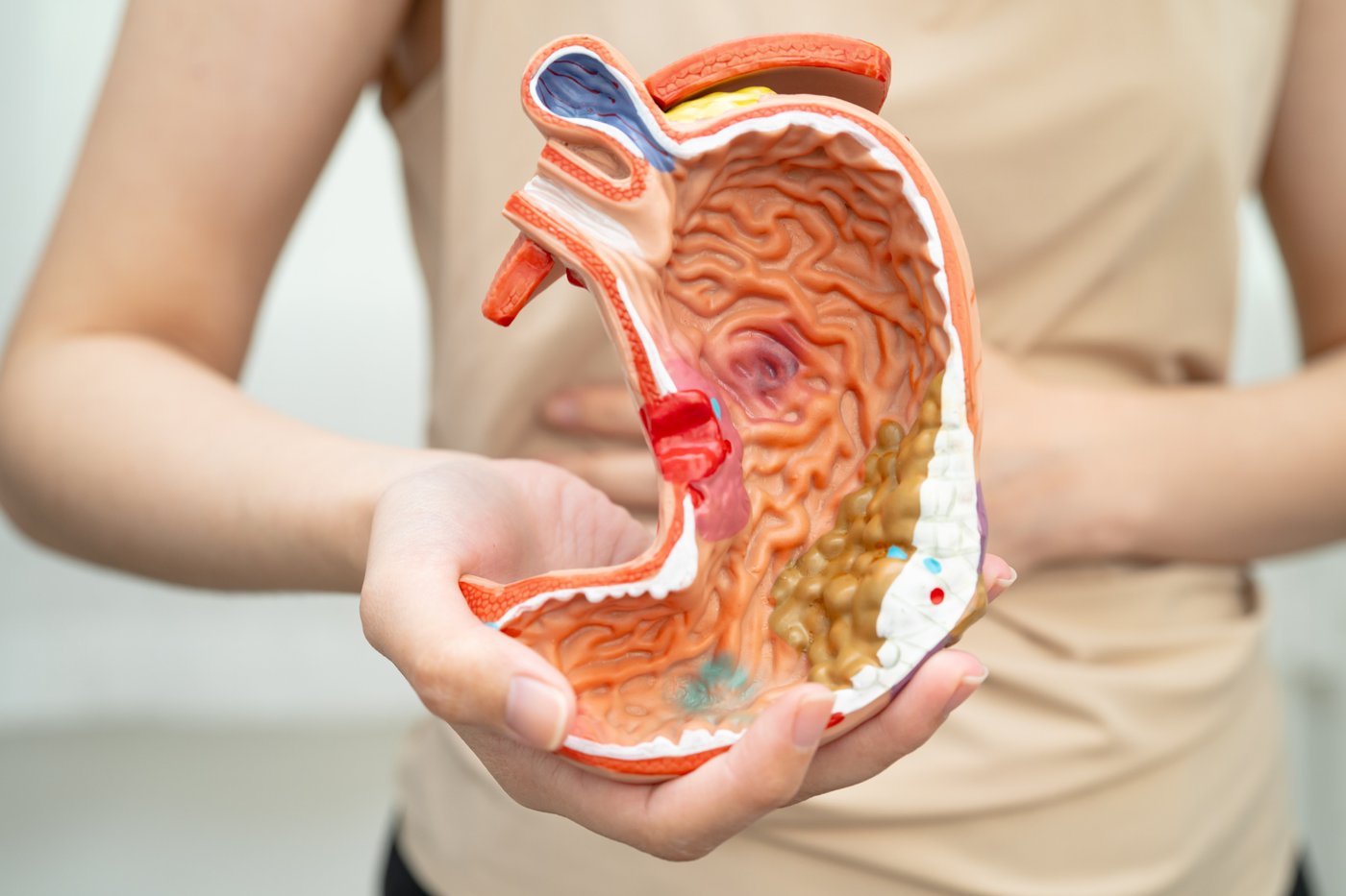
Your stomach is full of hydrochloric acid and enzymes designed to break down meat, which raises an obvious problem. Your stomach is itself made of tissue. Why does it not digest its own walls?
The answer is a defense system running around the clock. The stomach lining secretes a thick layer of mucus rich in bicarbonate, which neutralizes acid right at the surface of the tissue. This coating forms a protective gel between the harsh contents of the stomach and the delicate cells underneath. Below it sits a layer of epithelial cells packed tightly together to block acid from seeping through.
Even with that shield, some cells still take damage. So the stomach solves the problem with speed. The cells lining its inner surface are among the fastest-replaced in the body, turning over every few days. Damaged cells are shed into the stomach and swept away, while fresh cells produced deeper in the lining migrate up to take their place. The surface you have now is not the one you had last week.
This constant renewal is why the stomach can tolerate an environment that would burn most other tissue. It is also why the system is fragile in a specific way. When the mucus barrier breaks down — from a bacterial infection with Helicobacter pylori, or from heavy use of certain painkillers that suppress protective compounds — acid reaches the wall and eats into it. The result is a peptic ulcer.
The stomach's chemistry is worth respecting on its own terms. Its acid keeps the contents at a pH low enough to kill many swallowed pathogens and to activate the enzyme pepsin, which chops proteins apart. That same power is why the barrier and the rapid cell turnover are not optional extras. They are the only reason the organ does not consume itself.
Your skeleton is torn down and rebuilt for your whole life
Credit: Michael Heise / Unsplash
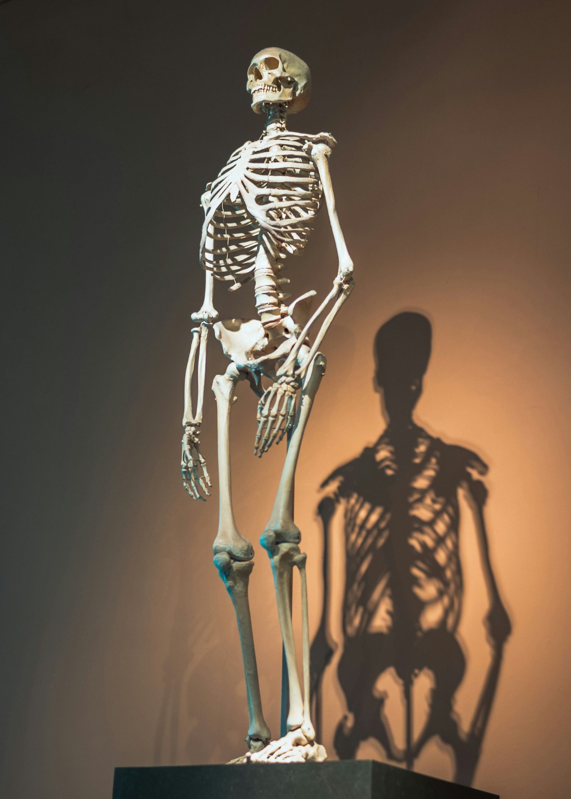
A skeleton looks like the most permanent thing in the body — dry, hard, unchanging, the part that survives long after everything else is gone. Living bone is nothing like that. It is active tissue, laced with blood vessels and nerves, and it is demolished and rebuilt continuously from birth to death.
Two types of cells run this process. Osteoclasts break down old bone, dissolving the mineral and releasing its calcium into the bloodstream. Osteoblasts follow behind, laying down fresh bone matrix that then hardens. This cycle, called remodeling, repairs tiny cracks from daily stress and lets the skeleton reshape itself in response to load. Bones you use heavily grow denser. Bones you stop using lose mass, which is why astronauts in weightlessness and people confined to bed rest shed bone quickly.
The pace is slow but relentless. A common estimate is that adults replace a meaningful fraction of their skeleton each year, so that much of the bone in your body is substantially renewed over the course of about a decade. The femur you have at 40 is largely not the same material you carried at 30.
This system does double duty. Beyond structural repair, it manages the body's calcium supply. Calcium is essential for nerve signaling, muscle contraction, and blood clotting, and the blood must hold it within a narrow range. When levels drop, hormones prompt osteoclasts to release calcium from bone. The skeleton acts as a mineral bank.
The balance can tip. If breakdown outpaces rebuilding over years, bones grow porous and brittle, the condition known as osteoporosis. Weight-bearing exercise, adequate calcium, and vitamin D all push the balance back toward construction. The takeaway is that your frame is not scaffolding bolted into place at adulthood. It is a tissue under lifelong construction, quietly rewriting itself while you go about your day.
You have a blind spot your brain paints over
Credit: Marc Schulte / Unsplash
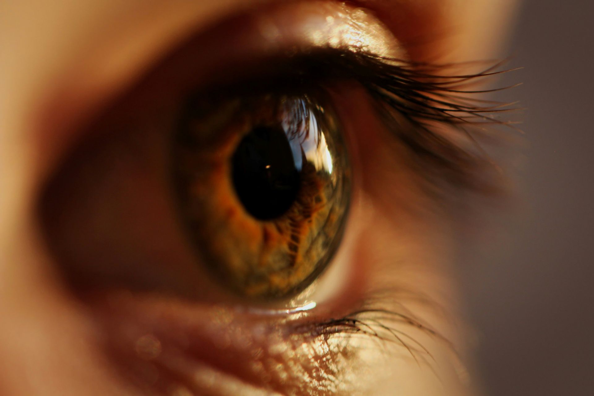
Every human eye has a hole in its vision, and almost no one notices. At the back of each eye, the optic nerve gathers the signals from millions of light-sensing cells and exits toward the brain. At the exact point where it leaves, called the optic disc, there is no room for photoreceptors. That patch of the retina is blind.
You can prove it to yourself. Close your right eye, hold up your left thumb and a fingertip a short distance apart, stare at the thumb, and slowly move the fingertip outward. At a certain angle the fingertip vanishes, then reappears. It has crossed the blind spot of your open eye.
In daily life the gap is invisible for two reasons. First, your eyes point in slightly different directions, so the blind spot of one eye covers a region the other eye can see. With both eyes open, the missing patches never overlap. Second, and stranger, your brain does not leave a hole even when you use one eye alone. It fills the gap by guessing what belongs there, based on the surrounding pattern, texture, and color. If you stare at striped wallpaper with one eye, the blind spot fills in with stripes.
This filling-in reveals something important about vision. You do not see the raw image landing on your retina. You see a version your brain constructs, edited to look complete and continuous. The blind spot is simply the clearest case where the construction becomes visible.
The same machinery smooths over the shadows cast by blood vessels sitting in front of your photoreceptors, and it stitches together the jerky snapshots your eyes take as they dart around a scene. What feels like a seamless window onto the world is closer to a running interpretation, and the blind spot is the seam the brain works hardest to hide.
You are taller in the morning than at night

Credit: Canva Images
Measure your height right after waking, then again just before bed, and the second number will usually be smaller. Most people lose around one to two centimeters of height over the course of a day. By morning, after a night lying down, it comes back.
The reason sits in your spine. Between each pair of vertebrae is an intervertebral disc, a cushion with a tough outer ring and a soft, water-rich core. These discs absorb shock and let the spine bend. When you stand, sit, and move through the day, gravity and body weight press down on the column, and the discs slowly squeeze out some of their water content. Compressed and slightly flattened, they take up less space, and the spine shortens.
At night the load comes off. Lying horizontal, the discs are no longer being pressed by your body weight, and they draw water back in, swelling to their full thickness. By the time you wake, the spine has recovered its length. The cycle repeats every day of your life.
The effect is more than a curiosity. Astronauts grow noticeably taller in orbit, sometimes by several centimeters, because the near absence of gravity lets their discs and spine expand without the usual compression. The extra height often brings back pain, and it reverses once they return to Earth.
Age changes the picture too. Over decades, the discs gradually lose water-holding capacity and thin out permanently, which is one reason people tend to shrink slightly as they get older. That long-term loss is separate from the daily rise and fall, but it comes from the same tissue.
The practical lesson is small but real. If you want a consistent height measurement, take it at the same time of day. Your body is not a fixed length. It is a structure that settles under load and lifts when the load is gone.
Your brain runs on a fifth of your energy
Credit: Natasha Connell / Unsplash
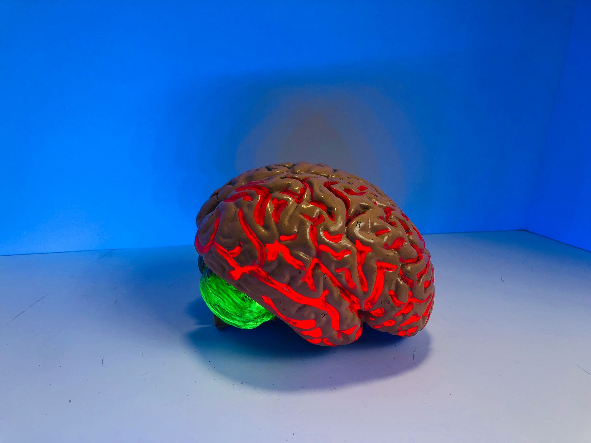
The brain accounts for roughly 2% of body weight in an adult, yet it consumes about 20% of the body's energy at rest. No other organ demands so much fuel relative to its size. A three-pound structure quietly burns a fifth of everything you take in.
That energy goes almost entirely to running neurons. Brain cells communicate through electrical signals, and maintaining the charge difference across their membranes is expensive. Every time a neuron fires, ions rush across its membrane, and the cell must then pump them back to reset for the next signal. This pumping runs constantly, across tens of billions of neurons, and it never fully stops. Even in deep sleep the brain keeps drawing heavily on glucose and oxygen.
The brain has almost no fuel reserves of its own. Muscles and the liver store energy as glycogen, but the brain holds very little. It depends on a steady delivery of glucose through the bloodstream, minute by minute. Cut off that supply and function fails within seconds, which is why fainting from a drop in blood flow happens so fast.
This hunger shapes human biology in deep ways. The energy cost of a large brain is thought to be one reason human evolution favored calorie-dense foods and cooking, which unlocks more energy from the same ingredients. It is also why the brain is so vulnerable. A stroke, which blocks blood flow to part of the brain, starves those neurons of fuel and oxygen and can kill them within minutes.
The demand does not scale much with mental effort in the way people imagine. Hard thinking raises energy use in specific regions only slightly. The baseline cost of simply keeping the machinery online dominates. Your brain is not expensive because you think hard. It is expensive because staying ready to think at all requires burning fuel around the clock.
You cannot tickle yourself
Credit: ROCKETMANN TEAM / Pexels
Try to tickle your own ribs or the soles of your feet, and it does not work. Someone else doing exactly the same thing can make you squirm. The failure is not about willpower or mood. It comes down to how your brain predicts your own movements.
Whenever you move, your brain sends a copy of the motor command to other regions, essentially a prediction of what your body is about to do and what sensation should follow. The cerebellum, a structure at the back of the brain, compares this prediction against the actual sensory signals coming in. When they match, the brain treats the sensation as self-generated and dampens it. This is why you do not startle at the feeling of your own footsteps or the movement of your own hand.
Tickling depends on surprise and unpredictability, so a self-generated touch, fully predicted in advance, gets muted before it can register as ticklish. Neuroscientist Sarah-Jayne Blakemore and colleagues demonstrated this using a device that let people move a lever to stroke their own palm. When the touch was delivered exactly as the person moved, it felt weak. When the researchers added a small delay or a change in direction between the movement and the touch, the prediction no longer matched, and the sensation felt more ticklish again.
This prediction system is not a quirk limited to tickling. It runs constantly, letting you tell the difference between sensations you cause and sensations the world imposes on you. It stabilizes your vision as your eyes move, and it lets you hold a cup without being distracted by the feeling of your own grip.
When the system breaks down, the consequences are serious. Some symptoms in conditions such as schizophrenia are linked to a failure to correctly flag self-generated actions, so a person's own inner speech or movement can feel as though it comes from outside. The inability to tickle yourself is one visible edge of a mechanism that helps define the boundary of the self.
Your nostrils work in shifts
Credit: Zulfugar Karimov / Unsplash
Breathe in and pay attention, and you may notice that one nostril is passing more air than the other. This is not a sign of a cold. For most people, most of the time, the two nostrils are not equally open, and which one leads changes throughout the day. The pattern is called the nasal cycle.
Inside each nostril, spongy tissue called the turbinates can swell or shrink as blood flows into or out of them. The autonomic nervous system, the same network that controls heart rate and digestion without conscious effort, alternates which side is congested. One nostril's tissue swells and restricts airflow while the other's shrinks and opens up. Then, over a period that varies but often runs a few hours, they switch.
Not everyone notices, and not every person shows a strong cycle, but it is a normal feature of nasal physiology rather than a defect. Researchers think the alternation gives each side of the nose a chance to rest and recover. The nasal lining works hard, warming and humidifying incoming air and trapping particles in mucus. Running one side at reduced flow may prevent it from drying out while the other side handles the bulk of breathing.
There is a smell-related angle too. Some odor molecules are detected best when they reach the receptors quickly, others when they linger. By having one nostril draw air fast and the other slow, the nose may sample a wider range of scents than a single uniform airflow would allow.
The cycle becomes obvious when you lie on your side, because the lower nostril often congests further. It is also why a mild cold can feel like it moves from one side of your nose to the other. The underlying cycle is always running. Illness simply makes its effects easier to feel. Your nose, it turns out, is quietly rotating its workload from one side to the other all day long.
Your red blood cells threw away their own DNA
Credit: Canva Images
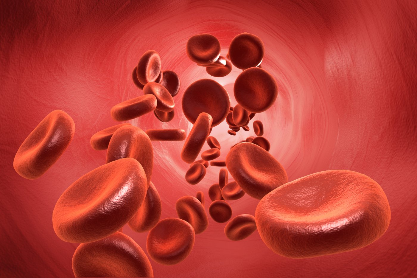
Nearly every cell in your body carries a nucleus holding a full copy of your DNA. Red blood cells are the great exception. As they mature, they eject their nucleus entirely, ending up as one of the few cell types in the body that carry no genetic blueprint at all.
The reason is efficiency. A red blood cell has one main job: to carry oxygen from the lungs to the rest of the body and ferry carbon dioxide back. It does this using hemoglobin, an iron-rich protein that binds oxygen. To pack in as much hemoglobin as possible, the cell clears out its internal structures during development. Out goes the nucleus, and out go the mitochondria and most other components. What remains is essentially a flexible bag of hemoglobin.
This design has real advantages. Without a bulky nucleus, the cell adopts its distinctive shape, a disc pinched in the middle on both sides, which maximizes surface area for gas exchange. The lack of internal machinery also lets the cell squeeze through the narrowest capillaries, some barely wider than the cell itself, bending and folding to pass through and then springing back.
There is a cost. Having discarded the equipment cells use to repair themselves and generate energy the usual way, a red blood cell cannot maintain itself indefinitely. It survives about 120 days in circulation before it wears out. The spleen and liver then remove the aging cells, and the body recycles their iron to build new ones. Your bone marrow produces millions of fresh red blood cells every second to keep pace.
The result is a cell that has traded longevity and self-repair for singular focus. It cannot divide, cannot fix damage, and carries none of the genetic information that defines the rest of your body. It is a specialist stripped down to a single purpose, and that ruthless simplification is exactly what makes it good at its job.
Your stomach acid is strong enough to harm your own tissue
Credit: Canva Images
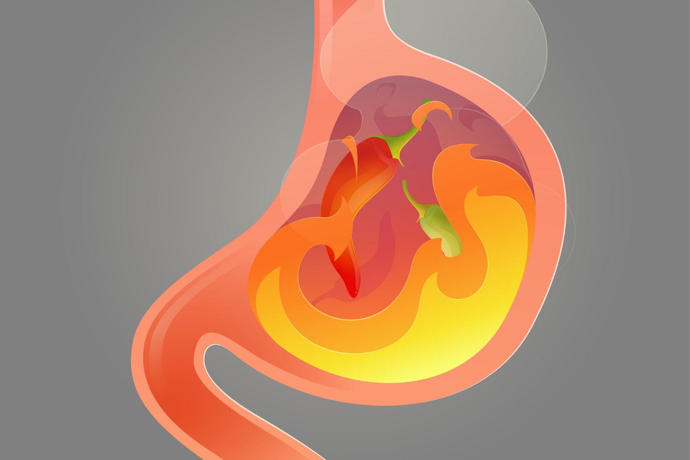
The acid your stomach produces to break down food is powerful by any measure. It is hydrochloric acid, the same compound used in industrial cleaning and metal processing, though at a lower concentration. In the stomach it drives the pH down to roughly 1.5 to 3.5, placing it among the most acidic environments in the body.
That acidity serves two purposes. It activates pepsin, an enzyme that dismantles proteins into smaller pieces the body can absorb, and it kills a large share of the bacteria and other pathogens riding in on your food. Many microbes that would sicken you never survive the trip through the stomach. The acid is a chemical checkpoint as much as a digestive tool.
At that strength, the acid can damage living tissue directly. This is why acid reflux is painful. When stomach contents rise into the esophagus, which lacks the stomach's heavy protective lining, the acid irritates and inflames the tissue, producing the burning feeling known as heartburn. Repeated exposure over years can cause lasting damage to the esophagus.
The stomach protects itself with the mucus and bicarbonate barrier described earlier, along with rapid cell replacement. When that defense holds, the acid does its work on food without touching the wall. When it fails, ulcers form.
The strength of the acid also explains a common piece of medical advice. Certain medications and supplements are absorbed differently depending on stomach acidity, and drugs that reduce acid production, taken by millions of people for reflux, can change how the stomach handles both food and microbes.
Popular claims that stomach acid can dissolve a razor blade overnight are exaggerated, but the underlying point holds. The fluid in your stomach right now is corrosive enough that, without a constantly maintained defense, it would begin to break down the very organ producing it. Digestion runs on a controlled chemical hazard sealed off just well enough to be useful.
Goosebumps are a leftover from a furrier past
Credit: Canva Images
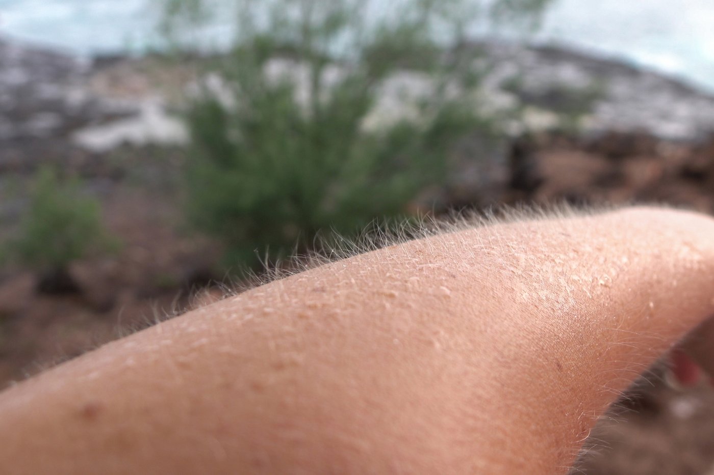
When you are cold or frightened, tiny bumps rise on your skin and the fine hairs stand up. The reaction is automatic. It comes from small muscles called arrector pili, each attached to the base of a hair follicle. When they contract, they pull the hair upright and pucker the surrounding skin into a bump. The trigger comes from the same nervous system that handles other involuntary responses to stress and temperature.
In humans the response does almost nothing useful, which is the point. It is a holdover from ancestors with dense body hair. In furry mammals, raising the hair serves two clear functions. In the cold, erect hairs trap a thicker layer of air against the skin, improving insulation. When threatened, a coat standing on end makes the animal look larger and more intimidating to a rival or predator. You can see both at work in a cat that puffs up when startled or a dog raising its hackles.
Humans kept the wiring but lost most of the fur, so the same reflex now produces a barely functional twitch of skin. It is one of the clearest examples of a vestigial trait, a feature retained from evolutionary history that no longer serves its original purpose in the current form of the animal.
The emotional trigger is worth noting. Goosebumps appear not only in the cold but during intense feeling, such as fear, awe, or the chill some people get from a piece of music. That connection reflects how the response is tied to the body's general stress and arousal system rather than to temperature alone.
Recent research has found that the same structures involved in goosebumps also play a role in signaling to the stem cells that regenerate hair, hinting that the muscle and follicle system has functions beyond the visible bump. Even so, the goosebump itself remains a small physical echo of a body plan we no longer have.
Your heart carries its own pacemaker
Credit: jesse orrico / Unsplash
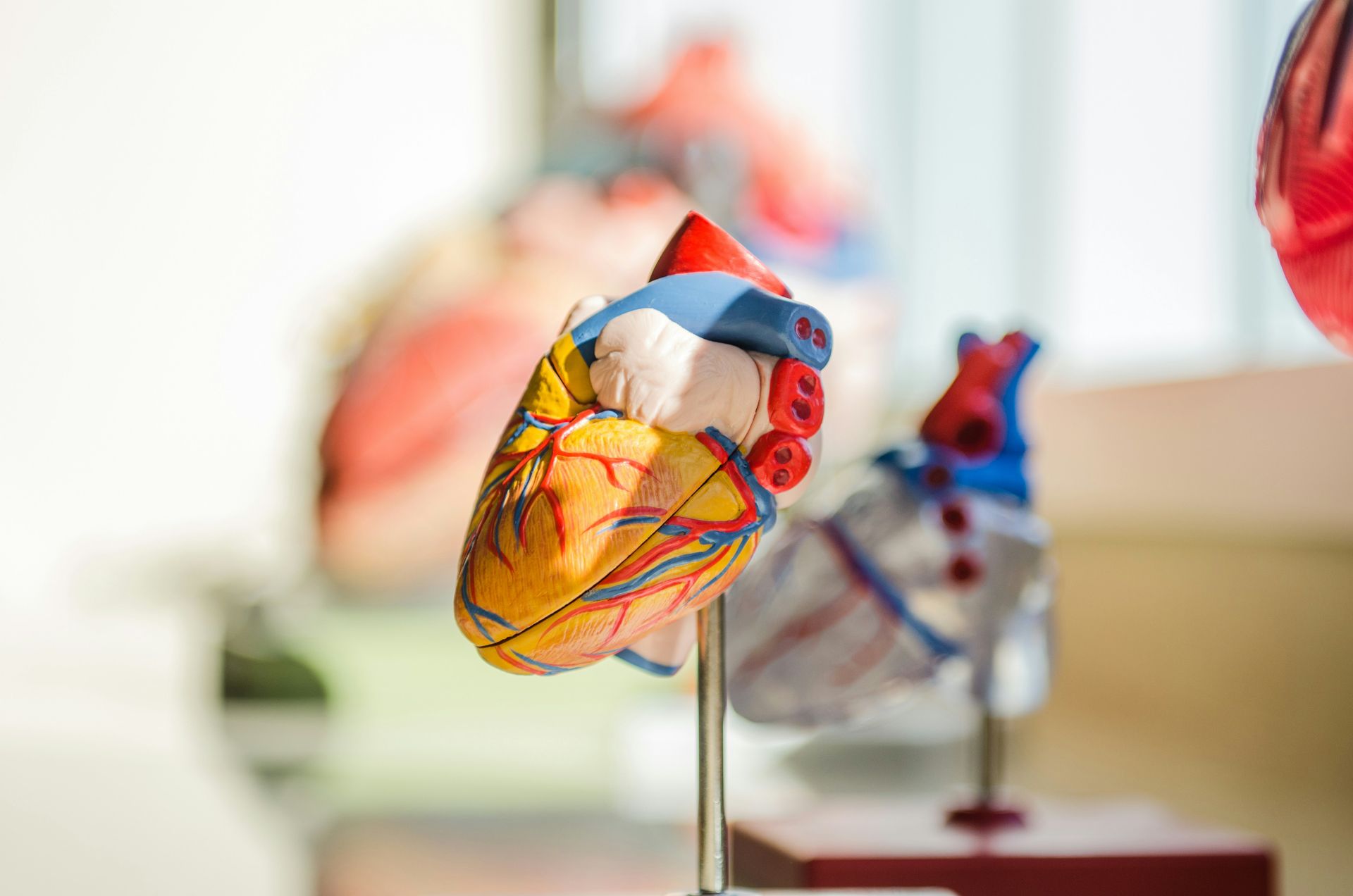
Most organs wait for instructions from the brain. The heart does not. It generates its own beat, using a built-in electrical system that fires whether or not the brain is involved. Remove a heart from the body and supply it with oxygen and nutrients, and it will keep beating on its own for a time. This is what makes heart transplants possible.
The rhythm starts in a small cluster of specialized cells called the sinoatrial node, sitting in the wall of the right upper chamber. These cells generate electrical impulses on their own, spontaneously and repeatedly, acting as the heart's natural pacemaker. Each impulse spreads across the heart in a coordinated wave, prompting the chambers to contract in the correct order so blood moves through in one direction.
The brain and nervous system do influence the heart, but as a regulator rather than a starter. Signals from the autonomic nervous system speed the heart up during exertion or fear and slow it down at rest. Hormones such as adrenaline push it faster. The heart's own node sets the baseline, and these outside signals adjust it up or down. Cut the nerve input and the heart still beats, just at a steadier default pace.
The heart also has a dense network of neurons embedded in its own walls, sometimes called the intrinsic cardiac nervous system or the heart's little brain. This local network helps fine-tune the beat and process signals without routing everything through the brain.
When the natural pacemaker fails or the electrical pathways misfire, the consequences are severe, from a dangerously slow rhythm to a chaotic one that cannot pump blood. Artificial pacemakers, small implanted devices that deliver electrical pulses, exist precisely to take over when the heart's own system falters.
The independence of the heartbeat is easy to overlook because it is so reliable. It runs on the order of 100,000 beats a day, for a lifetime, generating its own signal the entire time, without ever asking permission.
You were born with about 100 more bones than you have now

Credit: Jessica Donnelly / Pexels
Adults have 206 bones. A newborn has around 300. Growing up does not mean adding bones. It means losing them, as separate bones fuse together into single larger ones over the years of childhood and adolescence.
The extra count at birth reflects how the skeleton is built. Much of a baby's skeleton starts as cartilage, a softer, more flexible tissue that gradually turns to bone through a process called ossification. Some structures that will eventually become one solid bone begin as several separate pieces connected by cartilage. This flexibility serves a purpose. It lets the skull compress slightly to pass through the birth canal, and it gives the growing skeleton room to lengthen.
The skull shows this clearly. A newborn's skull is made of several plates separated by soft gaps called fontanelles, the soft spots parents are warned to protect. The gaps let the brain grow rapidly in early life. Over months and years the plates expand and knit together, closing the gaps and merging into the fused adult skull.
The spine and the pelvis follow similar paths. The sacrum, the wedge of bone at the base of the spine, forms from several vertebrae that fuse into one during adolescence and early adulthood. The three bones that make up each side of the pelvis also start separate and merge.
This is why bone age can be estimated from an X $TWTR-ray of a child's hand. The pattern of which bones have fused and how far the growth regions have closed reveals developmental age, sometimes more accurately than a birth certificate in certain contexts.
The fusion process is essentially complete by the mid-twenties, when the last growth regions close. At that point the count has settled at 206, though minor variation exists between individuals. The larger point is that skeletal development is a story of consolidation. You do not build a skeleton by gathering more parts. You build it by welding many small starting pieces into fewer, stronger, finished ones.
Your gut has its own nervous system
Credit: Canva Images
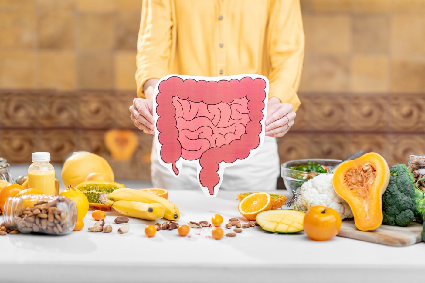
Your digestive tract does not need constant direction from the brain to do its job. Woven through the walls of the esophagus, stomach, and intestines is a vast network of neurons that can operate on its own. It is called the enteric nervous system, and it contains hundreds of millions of nerve cells, enough that some researchers describe it as a second brain.
This network controls the muscular contractions that move food along, coordinates the release of digestive enzymes, and regulates blood flow to the gut. It can run these processes without input from the brain or spinal cord, which is unusual. Most of the body's functions depend on signals routed through the central nervous system. The gut largely governs itself.
The enteric nervous system does communicate with the brain, along a two-way channel often called the gut-brain axis. The vagus nerve carries signals in both directions, and a large share of that traffic actually runs from the gut up to the brain rather than the other way around. The gut is constantly reporting on its state.
This connection helps explain sensations everyone has felt. Anxiety can produce nausea or an upset stomach. A serious digestive problem can affect mood and appetite. The gut also produces a large fraction of the body's serotonin, a chemical messenger involved in mood and, in the gut, in regulating movement of the intestines.
The microbiome adds another layer. The trillions of bacteria in the gut produce compounds that interact with this nervous system and with the signals traveling to the brain. Researchers are still working out how much these interactions shape mood, behavior, and disease, and the field remains active and unsettled.
What is not in doubt is the basic anatomy. Running the length of your digestive tract is a neural network large and capable enough to manage digestion largely on its own, quietly coordinating one of the body's most complex jobs while the brain attends to other things.
Your cornea has no blood supply
Credit: Rodrigo Gabotto / Pexels
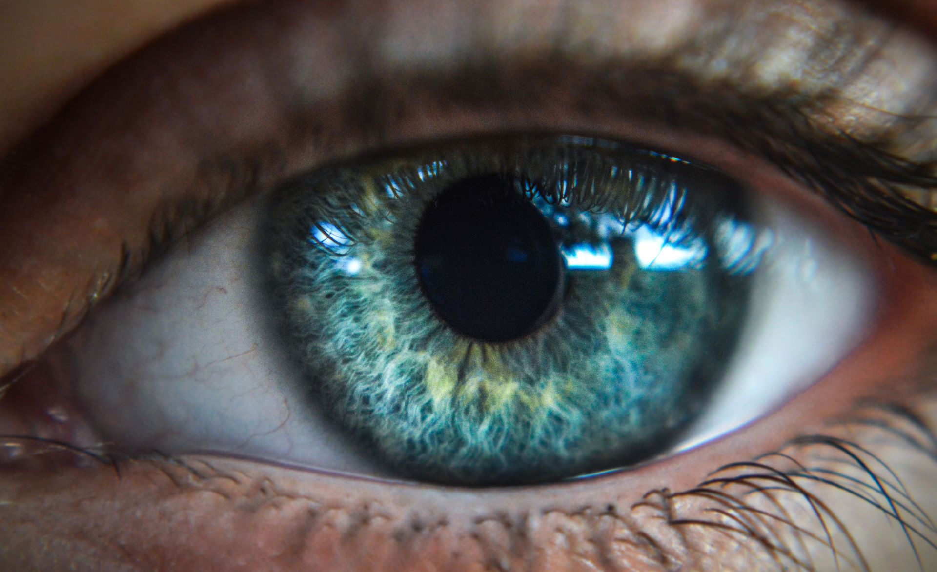
Almost every tissue in the body is fed by blood vessels delivering oxygen and nutrients. The cornea, the clear dome at the front of your eye, is a rare exception. It contains no blood vessels at all, and for a specific reason: blood vessels would block the light it needs to let through.
The cornea's job is to be transparent. It is the eye's outermost lens, bending incoming light and focusing it toward the retina. Any blood vessels running through it would scatter and absorb light, clouding vision. So the cornea evolved to stay clear by doing without the usual plumbing.
That leaves the problem of how it stays alive. The cornea gets its oxygen directly from the air. Oxygen dissolves into the tear film coating the eye's surface and passes into the corneal tissue. When you close your eyes, oxygen comes instead from small vessels in the eyelid and from the fluid inside the eye. Nutrients arrive from that internal fluid, called the aqueous humor, which bathes the back of the cornea.
This arrangement has practical consequences, especially for contact lens wearers. A lens sits directly on the cornea and can limit how much oxygen reaches it. Poorly fitting lenses, or lenses worn too long, can starve the cornea of oxygen and cause damage. Modern lens materials are designed specifically to let more oxygen pass through.
The lack of blood vessels has an upside in medicine. Because the cornea is not patrolled by the blood-borne immune cells that would normally attack foreign tissue, transplanted corneas are less likely to be rejected than most other transplanted organs. Corneal transplants are among the most common and successful transplant procedures, and donors do not need to be matched the way they are for organs like kidneys.
The clear window at the front of your eye, in other words, stays clear precisely because it gave up the blood supply that nearly every other part of you relies on to survive.
You inherit part of your DNA from only your mother
Credit: Sangharsh Lohakare / Unsplash
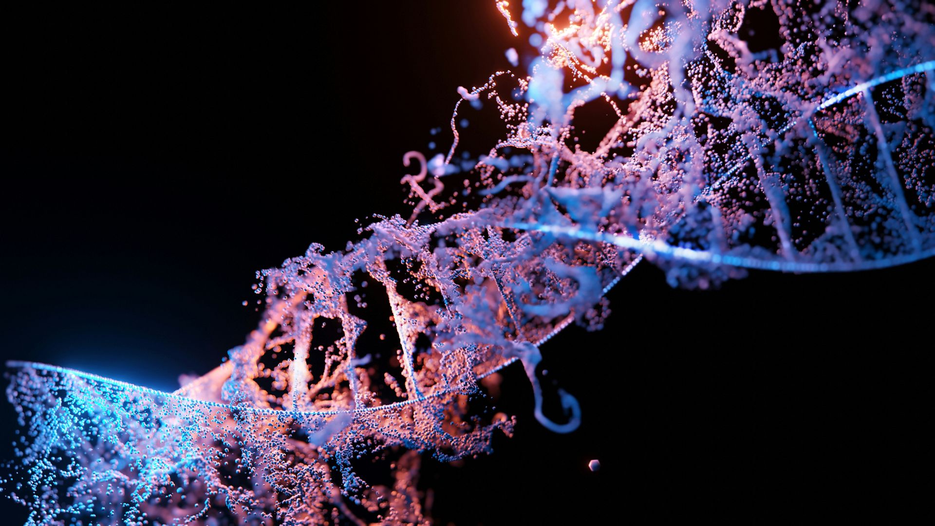
You carry two separate sets of genetic material. The famous one lives in the nucleus of your cells, drawn roughly equally from both parents. The second, smaller set lives outside the nucleus, inside the mitochondria, and it comes almost entirely from your mother.
Mitochondria are tiny structures inside nearly every cell, often described as the cell's power plants because they generate most of its usable energy. They are unusual in having their own small loop of DNA, separate from the DNA in the nucleus. This is a relic of their origins. Mitochondria are thought to descend from ancient bacteria that were absorbed by early cells billions of years ago and never left, keeping a remnant of their own genome.
When a sperm fertilizes an egg, it contributes nuclear DNA but almost none of its mitochondria. The egg supplies the mitochondria for the new cell, and any paternal mitochondria that do enter are typically destroyed. As a result, your mitochondrial DNA matches your mother's, which matches her mother's, tracing an unbroken maternal line back through generations.
This pattern makes mitochondrial DNA a powerful tool. Because it passes down the maternal line largely unchanged apart from slow mutations, scientists use it to trace ancestry and to study human migration across deep time. It is the basis for the concept of a most recent common maternal ancestor for all living humans.
It also matters for health. Mutations in mitochondrial DNA cause a distinct group of inherited diseases, and because the DNA comes from the mother, these conditions follow a maternal inheritance pattern that differs from ordinary genetic disease. This is the science behind so-called three-parent techniques, which aim to replace faulty mitochondria in an egg with healthy ones from a donor.
Rare cases of paternal mitochondrial inheritance have been reported, so the rule is not absolute. But for practical purposes, one slice of your genetic identity is a direct copy handed down along your mother's line alone.
Some of the dust in your home is made of you

Credit: Liana S / Unsplash
Look at a beam of sunlight in a still room and you will see fine particles drifting through it. A portion of that household dust is you, or rather the skin you have shed. Human skin renews itself constantly, casting off dead cells from its surface, and those flakes end up in the air and on every surface around you.
Your skin's outer layer, the epidermis, is in continuous turnover. New cells form at its base, gradually push toward the surface, flatten, die, and are shed. A person loses a large number of skin cells every hour, and the outermost layer of the epidermis is replaced roughly every month. Over a lifetime, that adds up to a considerable mass of discarded skin.
The popular claim that household dust is mostly dead skin is an exaggeration. Dust is a mix of many things, and its composition varies by home and setting. It includes fibers from clothing and furniture, soil and pollen tracked in from outside, tiny fragments of insects, and, in many homes, the microscopic remains and waste of dust mites. Human and pet skin is one ingredient among several, not the whole.
Dust mites are worth a mention because they connect directly to the shed skin. These microscopic relatives of spiders live in bedding, carpets, and upholstery, and they feed largely on flakes of human and animal skin. A used mattress and pillow can host large populations of them. Their waste is a common trigger for allergies and asthma, which is why washing bedding in hot water is standard advice for people with dust allergies.
None of this is a sign of a dirty home. Skin shedding is a normal, healthy process happening every second, on everyone, everywhere. The dust settling on your shelves is partly the ordinary residue of being alive in a body that is always quietly renewing its surface and leaving the old version behind.
Your taste buds are replaced every week or two
Credit: Canva Images
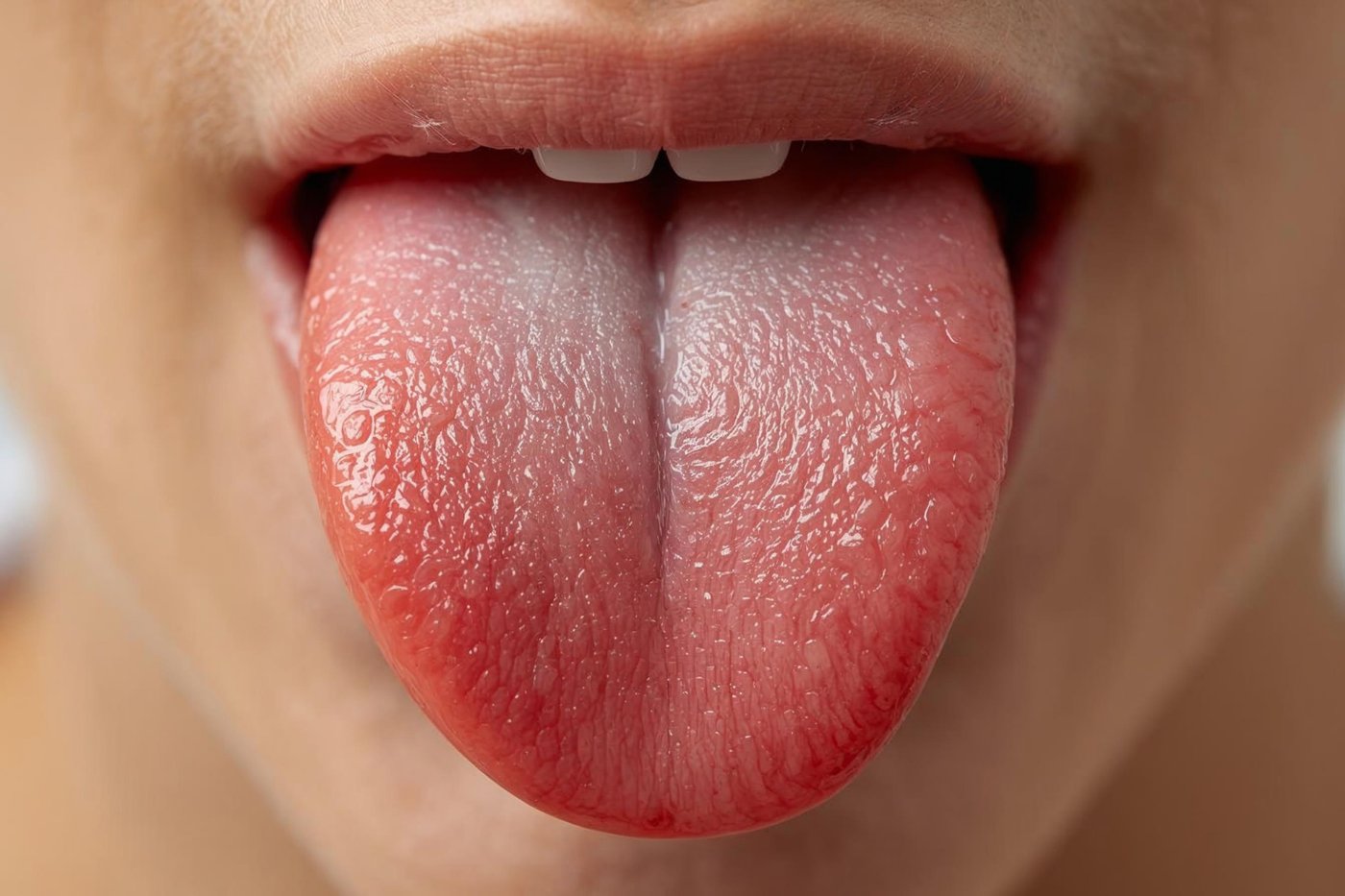
The taste buds that let you register sweet, salty, sour, bitter, and savory are not permanent fixtures. The cells that do the sensing are replaced continuously, with each generation lasting only a week or two before new cells take over. The tongue you taste with today is not made of the same sensing cells you had last month.
Taste buds are small clusters of cells, mostly housed in the bumps on your tongue called papillae, with others scattered on the roof of the mouth and in the throat. Within each bud sit the taste receptor cells that detect chemicals in food and drink and pass signals to the brain. These receptor cells work in a harsh environment, exposed to heat, cold, acid, rough textures, and a constant flow of food. Rapid replacement keeps the system functioning despite the wear.
This turnover explains a familiar experience. Burn your tongue on hot coffee or pizza and taste may dull in that spot for a few days, then return. The damaged cells are shed and replaced, and normal sensing resumes. The ability to recover from minor injury is a direct benefit of the constant renewal.
The common belief that different regions of the tongue are dedicated to different tastes, laid out in a fixed map, is a myth traced to a misreading of old research. Receptors for all the basic tastes are found across the tongue, not confined to zones.
Taste also changes over a lifetime as part of normal biology. Children tend to have more taste buds and often stronger reactions to bitter and strong flavors, which may be why many dislike foods like coffee or bitter greens that adults come to enjoy. The number and sensitivity of taste buds generally decline with age, which is one reason older adults sometimes find food blander and season it more heavily. Behind every meal is a sensing system rebuilding itself over and over.
Your body gives off a faint light
Credit: Canva Images
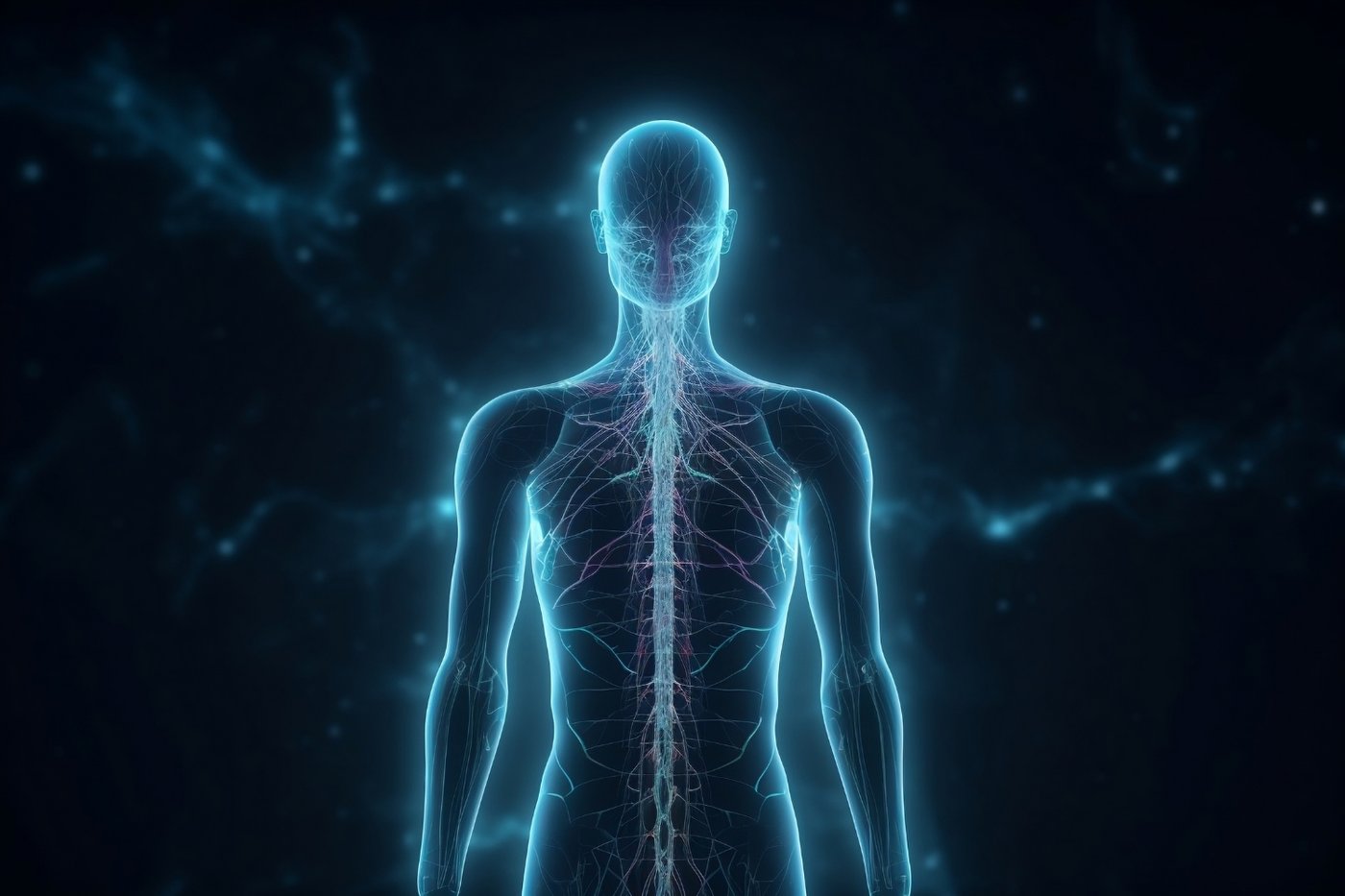
Humans emit light. Not the glow of a firefly or a deep-sea fish, but a genuine, measurable light given off by the body continuously. It is far too weak for the human eye to detect, roughly a thousand times fainter than the threshold of our vision, which is why no one has ever noticed it without instruments.
The phenomenon is called ultraweak photon emission, and it is a byproduct of ordinary metabolism. As cells carry out the chemical reactions that keep you alive, some of those reactions produce reactive molecules that release tiny amounts of light. The emission is not the same as the heat every warm body radiates in the infrared. It is visible-range light, just at an intensity so low it takes sensitive detectors in complete darkness to capture it.
A team of researchers in Japan, led by Masaki Kobayashi, documented this human glow in a 2009 study published in the journal PLOS ONE. Using an extremely sensitive camera in a light-sealed room, they imaged volunteers over several hours and found the whole body emits a faint light, with the glow rising and falling on a daily rhythm. The face tended to be brighter than the torso.
The intensity tracked with metabolic activity, which fits the idea that the light comes from the chemical work of living cells. The pattern followed the body clock, lowest in the morning and higher in the afternoon, consistent with how metabolism shifts across the day.
The finding is more than a curiosity. Because the emission reflects metabolic and chemical activity, researchers have explored whether measuring it could one day offer a non-invasive window into the body's internal state. For now the practical uses remain limited and the science continues.
The takeaway is that the human body is not a dark object. It is quietly luminous, giving off a whisper of light every moment you are alive, sealed away from notice only by how astonishingly faint it is.
Your appendix may be a safe house for gut bacteria
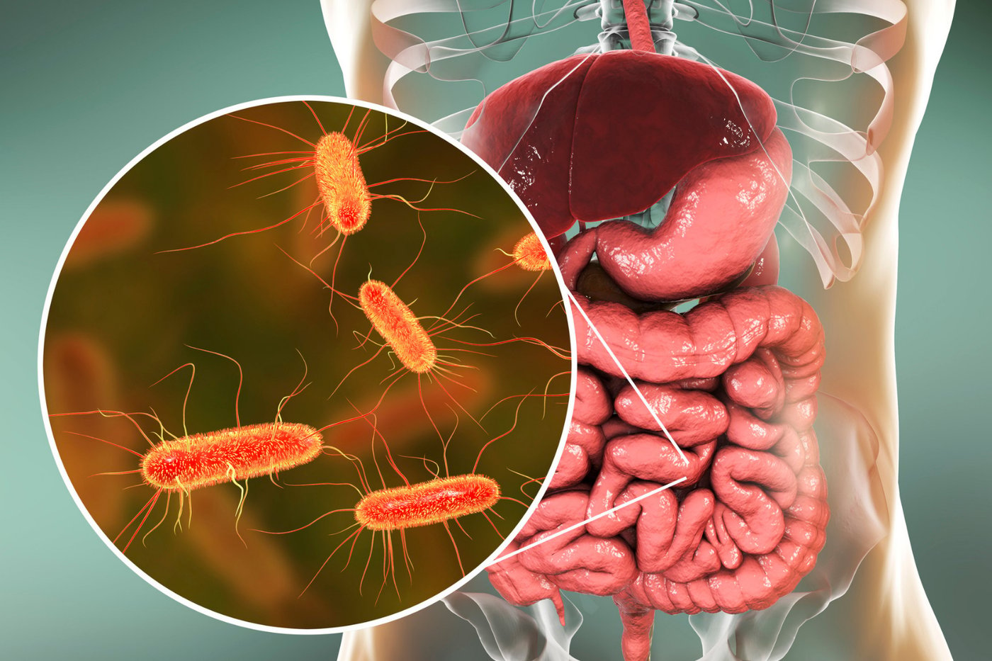
Credit: Canva Images
The appendix has long been treated as a useless leftover, a small dead-end pouch hanging off the large intestine whose only notable act is occasionally becoming inflamed and needing removal. People live normal lives without it, which reinforced the idea that it does nothing. That view is now under revision.
The leading modern hypothesis holds that the appendix serves as a reservoir, or safe house, for beneficial gut bacteria. Researchers at Duke $DUK University, including surgeon William Parker and colleagues, proposed in 2007 that the appendix stores a backup supply of the microbes that populate the intestines. Its location and shape support the idea. It sits below the main flow of the digestive tract in a spot where a protected community of bacteria could survive an event that flushes the rest of the gut.
The logic centers on recovery from illness. Severe diarrheal diseases, which have been common threats throughout human history, can strip the gut of its normal bacterial population. After the illness passes, the gut needs to be recolonized. A protected pocket of the right microbes, tucked away where the purge could not reach, would help restore a healthy community faster. In environments with frequent gut infections, that backup could have offered a real survival advantage.
The appendix is also rich in immune tissue, part of the network of lymphoid tissue associated with the gut. This fits the picture of an organ involved in managing the relationship between the body and its resident microbes, rather than an inert remnant.
The evidence remains a hypothesis rather than settled fact, and people who have had their appendix removed generally show no lasting harm, likely because modern sanitation and the ability of gut bacteria to recolonize from other sources make the backup less essential than it once was.
Still, the reversal is notable. An organ dismissed for generations as purposeless may in fact have played a quiet role in keeping the body's microbial partners alive through the worst that illness could do.