
20 things that happen inside your brain when meditation becomes a habit
The neuroscience of meditation has expanded significantly in the past two decades — and the findings are more qualified than most wellness content suggests

Thomas Benedetti / Pexels
Meditation research has a credibility problem that is not entirely its own fault. The genuine neuroscience of contemplative practice — a body of evidence built over two decades through neuroimaging studies, randomized controlled trials, and longitudinal research on long-term practitioners — has been systematically overstated by the wellness industry that has an interest in selling meditation as a cure for everything and understated by the skeptical reaction that this overselling provokes. Both distortions leave the person trying to understand what meditation actually does to the brain in a worse position than the evidence itself would put them.
The honest picture is this: meditation produces real, measurable, and in some cases substantial changes in brain structure, brain function, and the cognitive and emotional capacities those structures and functions support. It is not a cure for all conditions, its effects are not uniform across all practitioners or all meditation styles, and many of the most dramatic claims made in popular meditation content significantly exceed what the evidence supports. But the genuine findings are remarkable enough that they do not need to be exaggerated: structural changes in cortical thickness, reductions in amygdala reactivity, growth in hippocampal volume, improvements in attention network efficiency, reduction in default mode network activity — these are documented changes to the physical structure and functional organization of the brain, not placebo effects or self-report artifacts.
This list covers 20 of those findings, drawn from peer-reviewed neuroscience research. Each entry covers the specific finding, the mechanism behind it where known, the type and strength of the evidence, and the specific conditions under which the effect appears — because most meditation effects are not universal. They depend on the type of meditation practiced, the duration and consistency of practice, the individual's starting condition, and in some cases the quality of the training received. Where the evidence is preliminary or contested, this is noted. The goal is accuracy rather than inspiration — the honest account of what meditation does to the brain that the wellness industry rarely provides.
The default mode network quiets down

Abhayaranya Yoga Ashram / Pexels
The default mode network (DMN) — a set of brain regions including the medial prefrontal cortex, posterior cingulate cortex, and angular gyrus that is active during mind-wandering, self-referential thought, and rumination — shows reduced activity during meditation and reduced baseline activity in long-term meditators compared to non-meditators. This finding is one of the most consistently replicated in meditation neuroscience and is directly relevant to the subjective experience of meditation: the quieting of mind-wandering and self-referential thought that meditators describe corresponds to measurable reductions in DMN activation.
Judson Brewer's research at Brown University used real-time fMRI neurofeedback to document the specific DMN deactivation that experienced meditators produce, finding that experienced practitioners could more reliably and more completely deactivate the DMN than novices and that this skill transferred to non-meditative contexts — experienced meditators showed lower baseline DMN activity even when not meditating. The posterior cingulate cortex specifically, which acts as a hub of DMN activity, showed the largest reductions in activation in the most experienced practitioners.
The DMN finding is significant beyond the subjective experience of meditation: elevated DMN activity is associated with depression, anxiety, and the ruminative thinking patterns that characterize both conditions. The meditation-induced reduction in DMN activity is one of the proposed mechanisms through which regular meditation reduces depressive and anxious symptoms.
The amygdala becomes less reactive

cottonbro studio / Pexels
The amygdala — the brain's threat-detection and emotional-reactivity center — shows reduced gray matter volume and reduced reactivity to emotional stimuli in experienced meditators compared to non-meditators, and these changes have been documented both in long-term practitioners and in people who have completed as little as eight weeks of mindfulness-based stress reduction (MBSR).
Sara Lazar's research at Harvard was among the first to document structural amygdala changes from meditation: her 2005 study found that long-term meditators had significantly greater cortical thickness in regions associated with attention and interoception, and subsequent work from the same group and others documented the amygdala volume and reactivity changes that accompany these cortical changes. A 2010 study by Britta Hölzel and colleagues at Massachusetts General Hospital found that eight weeks of MBSR produced measurable reductions in amygdala gray matter density, corresponding to participants' self-reported reductions in stress.
The amygdala reactivity finding is clinically significant: reduced amygdala reactivity means that the emotional threat-response system activates less strongly in response to the same provocations, producing a more stable emotional baseline. This is not the same as emotional numbness — meditators show normal emotional responses to genuine threats — but a reduction in the hair-trigger reactivity that characterizes anxiety disorders and high-stress states.
Cortical thickness increases in attention regions
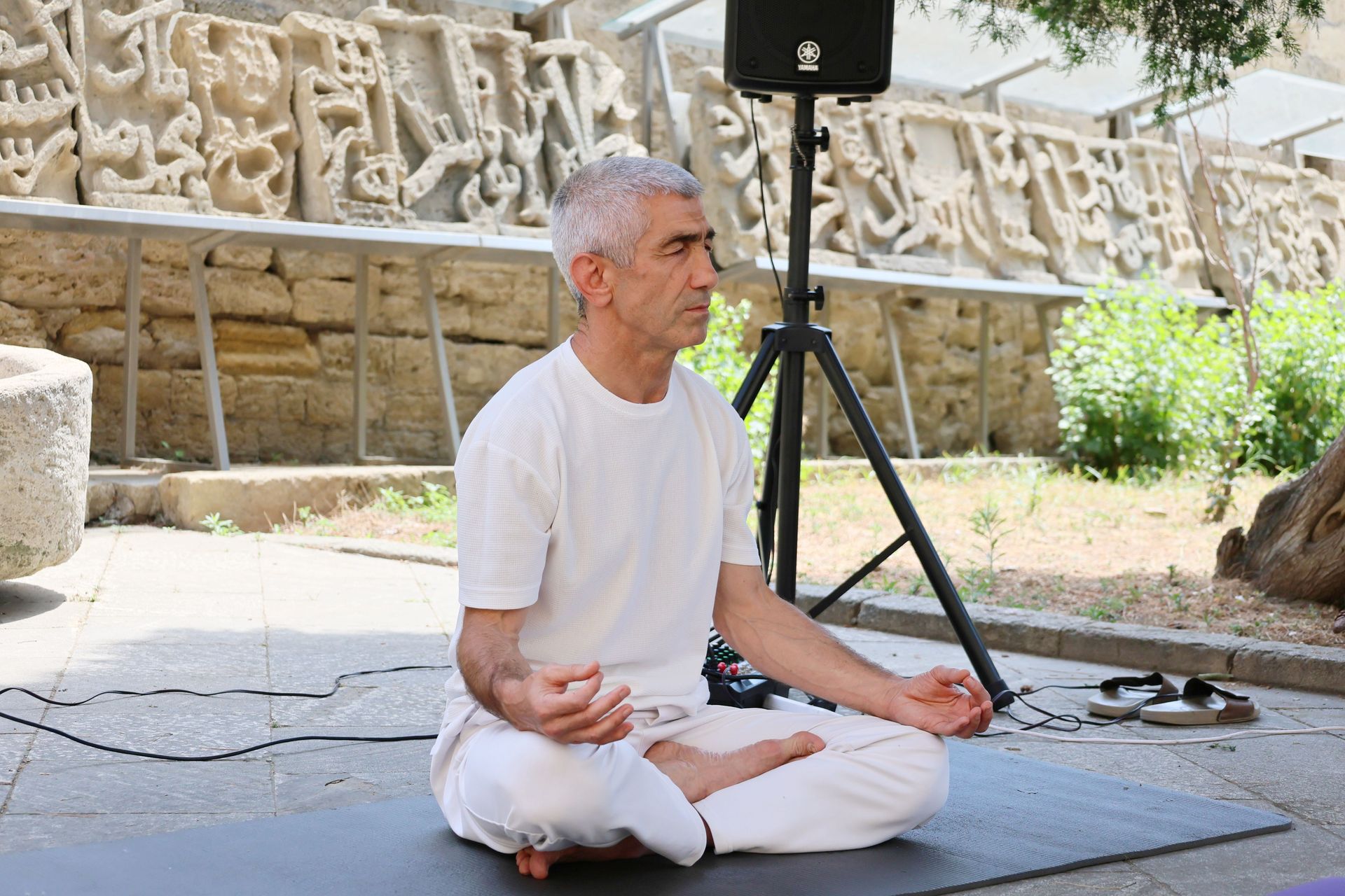
Tahir Xəlfəquliyev / Pexels
Regular meditation practice is associated with increased cortical thickness in brain regions involved in attention, interoception, and sensory processing — specifically the prefrontal cortex, the right anterior insula, and the right sensory cortex. This finding, documented in Lazar's 2005 study and replicated in multiple subsequent studies, suggests that meditation produces structural changes in the brain tissue associated with the cognitive capacities it trains, and that these changes are detectable as differences in the physical thickness of the cortex.
Cortical thickness normally decreases with age — the prefrontal cortex, which is the region most associated with attention regulation, planning, and executive function, thins progressively from early adulthood. The finding that long-term meditators show cortical thickness in these regions equivalent to people 5 to 10 years younger than their chronological age suggests that meditation may partially counteract the age-related cortical thinning in attention-related regions — though this interpretation requires caution, as the cross-sectional studies that established this finding cannot rule out the possibility that people with thicker cortex in these regions are more likely to sustain a meditation practice.
Longitudinal studies — which track the same individuals before and after beginning meditation practice — are more informative on this point, and several have documented measurable increases in cortical thickness after 8 to 12 weeks of regular practice, supporting the causal interpretation.
Hippocampal volume increases

Vitaly Gariev / Pexels
The hippocampus — the brain region most directly involved in the formation of new memories, spatial navigation, and the regulation of the stress response — shows greater gray matter volume in experienced meditators than in non-meditators, and longitudinal studies have documented hippocampal volume increases following MBSR training. The hippocampus is one of two brain regions known to produce new neurons in adults (hippocampal neurogenesis), and the volume increases from meditation are consistent with increased neurogenesis driven by the reduced cortisol and increased BDNF (brain-derived neurotrophic factor) that meditation produces.
The hippocampal finding is specifically important because hippocampal volume is reduced in major depressive disorder, PTSD, and chronic stress — three conditions for which meditation has the most consistent evidence of benefit. The mechanism connecting meditation to improved outcomes in these conditions likely runs through the hippocampus: reduced cortisol from the stress-reduction effect of meditation, increased BDNF from the neuroplasticity effects, and the resulting hippocampal volume increase that improves memory function and stress regulation.
Kirk Erickson's research group, which documented hippocampal volume increases from aerobic exercise, has also examined the hippocampal effects of meditation and found that the two interventions appear to affect hippocampal volume through overlapping but partially distinct pathways, suggesting that combining exercise and meditation may produce additive hippocampal benefits.
Attention networks become more efficient

Yan Krukau / Pexels
The three attention networks identified by Michael Posner and colleagues — the alerting network (which maintains a state of readiness), the orienting network (which selects specific information for focused attention), and the executive attention network (which resolves conflict between competing stimuli and maintains goal-directed attention) — all show measurable improvements in efficiency after meditation training, but the specific improvements differ by meditation style.
Focused attention meditation (FA), which trains the ability to sustain attention on a single object (typically the breath) and to notice and redirect mind-wandering, primarily improves the executive attention network — reducing the time required to detect attentional failures (mind-wandering) and to redirect attention back to the intended object. Open monitoring meditation (OM), which involves a non-reactive awareness of whatever arises in consciousness without directing attention to any specific object, primarily improves the alerting network — enhancing the readiness to notice novel stimuli.
Antoine Lutz's research at the University of Wisconsin, working with long-term meditators recruited through Richard Davidson's collaboration with the Dalai Lama, documented the attention network improvements in experienced practitioners using a combination of fMRI, EEG, and behavioral attention tasks, and found that the efficiency gains were associated with reduced neural effort — experienced meditators produced the same attention performance with less activation of the relevant brain networks, suggesting genuinely more efficient neural processing rather than simply increased effort.
The brain's response to pain changes

Kaboom Pics / Pexels
Regular meditation practice changes how the brain processes pain — not by reducing the intensity of the sensory signal but by reducing the emotional reactivity to that signal, decoupling the "unpleasantness" component of pain from its sensory intensity. This distinction — between how much something hurts (the sensory dimension) and how distressing it is (the affective dimension) — is one of the most important findings in pain neuroscience and one that meditation specifically addresses.
Nerve Fadel Zeidan's research at Wake Forest University used thermal pain stimulation in meditators and non-meditators with fMRI monitoring and found that meditation significantly reduced pain unpleasantness ratings (by approximately 57% in one study) while reducing pain intensity ratings somewhat less (approximately 40%). The neural correlates of this effect were changes in activity in the anterior cingulate cortex (which processes pain's emotional significance) and the orbitofrontal cortex (which integrates emotional and sensory information), rather than changes in the primary somatosensory cortex (which processes the raw sensory signal).
The practical implication is that meditation does not make pain disappear but changes the relationship to pain — the experience of pain as catastrophic, as requiring immediate escape, as dominating consciousness — which is precisely the dimension of pain that is most clinically relevant in chronic pain conditions where the sensory signal cannot be eliminated.
Gamma wave activity increases in experienced meditators

Vlada Karpovich / Pexels
Experienced meditators practicing compassion meditation show dramatically elevated gamma brainwave activity — synchronous neural oscillations at frequencies above 30 Hz — compared to both their own baseline states and to novice meditators. Richard Davidson's research with Tibetan Buddhist monks who had each accumulated 10,000 or more hours of meditation practice documented gamma oscillations of unusual amplitude and synchrony that had never previously been recorded from the human scalp.
Gamma oscillations are associated with the integration of information across distributed brain regions, with moments of cognitive insight, and with heightened conscious awareness. The specific gamma signature of experienced compassion meditation practice — which involves the cultivation of a pervasive sense of love and concern for all beings rather than focused attention on a specific object — is distinct from the gamma activity associated with other cognitive states and suggests a specific mode of brain organization that long-term meditation practice produces.
The novice versus expert comparison is important for interpreting this finding: the gamma elevation in experienced practitioners was not present in novices attempting the same compassion meditation, indicating that the effect is the product of thousands of hours of practice rather than the immediate effect of the meditation instruction. The specific neural changes required to produce this gamma signature appear to take years of consistent practice to develop.
Emotional regulation improves through prefrontal-amygdala connectivity

Mart Production / Pexels
The ability to regulate emotional responses — to modulate the intensity of an emotion, to reframe an emotional situation, to allow an emotion to arise and pass without being overwhelmed by it — depends on the functional connectivity between the prefrontal cortex (which exercises top-down regulation) and the amygdala (which generates the bottom-up emotional response). Regular meditation strengthens this connectivity, improving the efficiency of the top-down regulatory pathway and reducing the likelihood that amygdala-generated emotional responses will overwhelm prefrontal regulation.
This finding connects two separate lines of meditation research: the structural changes in the prefrontal cortex described in the cortical thickness entry and the amygdala reactivity changes described in the amygdala entry. Both changes contribute to improved emotional regulation, and they are connected functionally: a thicker, better-developed prefrontal cortex is better able to regulate a less reactive amygdala, and the combination produces emotional regulation improvements that exceed what either change alone would suggest.
Behavioral evidence for improved emotional regulation in meditators includes faster recovery from negative emotional stimuli, lower ratings of negative affect in response to aversive images, and reduced emotional reactivity in laboratory paradigms designed to provoke frustration. These behavioral changes map directly onto the neural changes in prefrontal-amygdala connectivity, providing convergent evidence for the mechanism.
The insula grows and becomes more active

Anil Sharma / Pexels
The insula — a cortical region buried within the lateral sulcus, involved in interoception (the perception of internal bodily states), empathy, and the integration of emotional and physical experience — shows both increased gray matter volume and increased functional activity in experienced meditators compared to non-meditators. The insula is the brain's primary interface between physical body states and conscious awareness, and its development through meditation practice corresponds to the increased body awareness, increased interoceptive sensitivity, and increased empathic resonance that meditators report.
The interoceptive function of the insula is directly relevant to several meditation practices: body scan meditation (systematic attention to physical sensations throughout the body) and breath awareness meditation (attending to the physical sensations of breathing) both directly train interoceptive attention, and the insula development that follows this training reflects the neural correlate of improved interoceptive skill. The right anterior insula specifically — which integrates interoceptive signals with emotional processing — shows the largest changes in meditators, consistent with meditation's specific effects on the emotional-interoceptive interface.
The empathy connection is also well-supported: the insula is one of the primary neural substrates of affective empathy (feeling what another person feels), and the insula development in meditators who practice compassion and loving-kindness meditation corresponds to increased empathic accuracy and increased prosocial behavior in behavioral measures.
Stress hormone levels fall over time

Tim Gouw / Pexels
Regular meditation practice reduces the baseline secretion of cortisol — the primary glucocorticoid stress hormone — and reduces the cortisol response to acute psychological stressors. These effects have been documented in multiple randomized controlled trials of MBSR and other meditation programs using salivary cortisol as an objective biological measure rather than relying solely on self-reported stress.
The mechanism of cortisol reduction from meditation involves the regulatory relationship between the prefrontal cortex, the hippocampus, and the HPA axis (the hypothalamic-pituitary-adrenal axis that controls cortisol secretion). The prefrontal cortex and hippocampus both contain glucocorticoid receptors and both provide inhibitory input to the HPA axis — when either region is stimulated, it reduces HPA axis output and therefore cortisol secretion. The meditation-induced development of prefrontal cortex thickness and hippocampal volume described in earlier entries corresponds to a strengthened inhibitory input to the HPA axis and therefore reduced cortisol output.
The practical significance extends beyond the subjective experience of stress reduction: chronically elevated cortisol accelerates biological aging through multiple mechanisms (telomere shortening, hippocampal damage, inflammatory activation), and the meditation-induced reduction in cortisol baseline is one of the primary mechanisms through which long-term meditation practice may produce the longevity-related benefits that have been documented in meditating populations.
White matter integrity improves

Kaboom Pics / Pexels
White matter — the myelinated axonal tracts that connect different brain regions and allow rapid communication between them — shows improved structural integrity in experienced meditators on diffusion tensor imaging (DTI) measures, particularly in the anterior cingulate cortex and the tracts connecting frontal regions to limbic structures. Improved white matter integrity indicates better-insulated, faster-conducting neural connections between the brain regions involved in attention regulation and emotional control.
Yi-Yuan Tang's research at Texas Tech University documented white matter changes in the anterior cingulate cortex region after as little as 11 hours of integrative body-mind training (IBMT), a specific mindfulness-based training protocol. The anterior cingulate cortex is a key node in the executive attention network, and the white matter changes in this region correspond to the attention improvements documented in behavioral studies after IBMT.
The mechanism of white matter improvement from meditation likely involves both increased myelination (the fatty sheath around axons that improves conduction speed and efficiency) and increased axonal density in the relevant tracts. The speed of these changes — documented after only 11 hours of training in Tang's studies — suggests that some of the white matter effects reflect rapid experience-dependent plasticity rather than the slower processes of myelination, though the long-term picture may involve both.
Mind-wandering decreases

Михаил Крамор / Pexels
The tendency toward mind-wandering — the spontaneous, unbidden departure of attention from the current task or intended focus — is one of the most reliably reduced phenomena in meditation research, both during meditation itself and in everyday cognitive tasks assessed outside of meditation sessions. A large experience-sampling study by Matthew Killingsworth and Daniel Gilbert at Harvard, which tracked people's thoughts throughout the day using smartphone prompts, found that people's minds wandered approximately 47% of the time during daily activities — and that mind-wandering was associated with reduced happiness regardless of what people were doing when it occurred.
Meditation specifically trains the capacity to detect mind-wandering (metacognitive awareness) and to redirect attention, and research using experience sampling methods similar to Killingsworth and Gilbert's has found that experienced meditators show significantly lower rates of mind-wandering in everyday life than non-meditators. The neural correlate is the reduced default mode network activity described in the first entry — mind-wandering is the subjective experience of DMN activation, and the meditation-induced DMN reduction corresponds directly to reduced mind-wandering.
The reduction in mind-wandering from meditation has downstream effects on both happiness (consistent with Killingsworth and Gilbert's finding that mind-wandering produces unhappiness) and cognitive performance (mind-wandering during tasks impairs performance, and its reduction correspondingly improves it).
Compassion and empathy circuits strengthen

Jsme MILA / Pexels
Compassion meditation — specifically loving-kindness meditation (metta bhavana) and related practices that involve the deliberate cultivation of warm regard for oneself and others — produces measurable increases in activity and connectivity in the neural circuits associated with empathy, compassion, and prosocial motivation. These circuits include the insula (for affective empathy, described in the insula entry), the anterior cingulate cortex (for the integration of empathic information with behavioral response), and the medial orbitofrontal cortex (for the positive valuation that underlies compassionate motivation).
Tania Singer's research group at the Max Planck Institute documented the neural distinction between empathy (sharing another's emotional state) and compassion (the motivated concern for another's wellbeing that does not require sharing their suffering), and demonstrated that compassion meditation specifically trains the compassion circuit rather than simply amplifying empathy — an important distinction because excessive empathy without compassion can produce emotional exhaustion (empathic distress), while compassion provides the motivational basis for helping behavior without the burnout.
The behavioral evidence for compassion increases from meditation training includes increased prosocial behavior in economic games, increased willingness to help strangers in staged situations, and increased self-reported compassion for both self and others. These behavioral changes are associated with the neural changes in the compassion circuit, providing convergent evidence for a genuine compassion increase rather than a social desirability bias in self-report.
Neuroplasticity markers improve

Katrin Bolovtsova / Pexels
BDNF (brain-derived neurotrophic factor) — the primary molecular signal for neuroplasticity, supporting the survival of existing neurons, promoting the growth of new ones, and mediating synaptic strengthening — is elevated in regular meditators compared to non-meditators and shows acute increases following meditation sessions. BDNF is the molecular mechanism through which many of the structural brain changes described in this list are produced: the hippocampal neurogenesis, the cortical thickening, the synaptic strengthening in attention networks all require BDNF signaling.
The elevation of BDNF from meditation overlaps with the elevation from aerobic exercise — both interventions produce similar acute increases in circulating BDNF, and both produce similar structural brain changes in the hippocampus and prefrontal cortex. The similarity suggests a partially shared mechanism: both exercise and meditation reduce cortisol (which inhibits BDNF), increase BDNF directly, and stimulate the BDNF-dependent neuroplastic processes that produce structural changes.
The BDNF connection is significant for understanding why meditation's brain benefits extend beyond stress reduction: the same molecular pathway that exercise uses to build a healthier brain is activated by meditation, suggesting that the two practices may be addressing overlapping aspects of brain health through a shared mechanism.
Theta wave activity increases during meditation

Vitaly Gariev / Pexels
Theta brainwave activity — oscillations at 4 to 8 Hz — increases during meditation, particularly during the specific transitional state between waking and sleep (the hypnagogic state) that some deep meditation practitioners deliberately induce and maintain. Theta waves are associated with creative insight, memory consolidation, and the relaxed, receptive state of awareness that characterizes successful meditation, and their increase during practice reflects the specific neural state that meditation produces.
The increase in frontal theta activity during focused attention meditation has been documented in multiple EEG studies and is one of the most reliable neural signatures of meditative absorption. It is distinct from the theta activity associated with drowsiness or sleep onset — meditators show frontal theta with maintained alertness, a combination that is uncommon in non-meditating individuals and appears to require training to produce reliably.
Long-term meditators show higher baseline frontal theta activity even outside of formal meditation sessions, suggesting that the meditative neural state leaves a lasting signature on resting brain activity. This resting theta elevation may correspond to the quality of relaxed, open awareness that experienced meditators describe as their ordinary baseline state rather than a special meditative achievement.
The brain ages more slowly in long-term meditators

Gustavo Fring / Pexels
Multiple neuroimaging studies have found that long-term meditators show less age-related brain atrophy than non-meditators — specifically, the cortical thinning and gray matter reduction that normally accompany aging are less pronounced in people who have meditated regularly for years or decades. A 2015 study by Florian Kurth and colleagues found that long-term meditators in their 50s had gray matter volumes comparable to non-meditating adults in their 20s to 30s in multiple brain regions.
The mechanism of this apparent neuroprotective effect likely involves the combination of reduced cortisol (which directly causes hippocampal and prefrontal atrophy when chronically elevated), increased BDNF (which supports neuronal survival and opposes age-related neurodegeneration), and the maintenance of active attention networks that provides the cognitive stimulation associated with slower cognitive aging.
The caution required in interpreting this finding is the direction of causality: the cross-sectional studies that document the brain age difference cannot rule out the possibility that people with younger-looking brains are more likely to sustain a long-term meditation practice, rather than the meditation causing the younger-looking brain. Longitudinal studies tracking brain aging in meditators versus non-meditators over years are the necessary evidence, and while several are underway, the long timeframes required mean that this specific finding remains more suggestive than definitive.
Focus and concentration improve measurably

Jsme MILA / Pexels
The ability to sustain attention on a chosen object — measured by behavioral tasks including the attention network task, the sustained attention to response task, and the flanker task — improves significantly after meditation training across multiple randomized controlled trials. These are objective behavioral measures rather than self-report, and their improvement indicates genuine changes in attentional function rather than simply the belief that one is more focused.
The concentration improvements are most pronounced for focused attention meditation (FA) — the practice of sustained attention to a single object with redirection when mind-wandering occurs — which directly trains the attentional skill being measured. The magnitude of improvement depends substantially on practice duration: studies using 8-week MBSR programs show modest but consistent improvements, while studies of experienced practitioners with thousands of hours of practice show much larger effects that approach the performance of the most focused non-meditating individuals.
Importantly, the attention improvements from meditation appear to transfer across tasks — meditators do not simply become better at the specific tasks practiced during meditation but show improved attention in novel attentional tasks as well, indicating that the improvement reflects a genuine capacity enhancement rather than task-specific learning.
Sleep quality improves through brain changes

Miriam Alonso / Pexels
Regular meditation practice improves sleep quality through specific changes to the brain's sleep-wake regulatory systems rather than simply through the general stress-reduction effects that improve sleep in non-meditators. Mindfulness-based therapy for insomnia (MBTI) — a specific meditation-based treatment program — has demonstrated efficacy in multiple randomized controlled trials for both sleep onset latency (time to fall asleep) and wake after sleep onset (time awake during the night), with effect sizes comparable to cognitive behavioral therapy for insomnia (CBT-I), the current gold standard treatment.
The neural mechanism of meditation's sleep benefit involves the reduction in pre-sleep cortical arousal — the elevated brain activity that prevents sleep onset in insomnia — through the prefrontal regulation of the arousal systems that meditation strengthens. EEG studies of meditators show increased pre-sleep theta activity and reduced high-frequency beta activity (associated with cognitive arousal) in the period before sleep onset, consistent with the subjective experience of falling asleep more easily and the behavioral improvement in sleep onset latency.
Long-term meditators also show improvements in sleep architecture — specifically increased slow-wave sleep percentage — that go beyond what the stress-reduction effect alone would predict, suggesting that the broader brain changes from meditation practice (the cortical thickening, the improved prefrontal regulation, the DMN quieting) contribute to sleep quality improvements that accumulate with sustained practice.
Sense of self becomes less fixed

Blue Bird / Pexels
One of the most philosophically interesting findings in contemplative neuroscience is the change in self-referential processing that long-term meditation practice produces. The narrative self — the sense of being a continuous, consistent entity with a coherent autobiographical story — is associated with DMN activity, and the meditation-induced reduction in DMN activity corresponds to a loosening of the fixed sense of self that characterizes most people's ordinary experience.
Advanced meditators practicing non-self or insight meditation — practices specifically designed to investigate the nature of the self — show further reductions in self-referential processing than those practicing focused attention or stress-reduction meditation, consistent with the specific cognitive objective of these practices. Judson Brewer's research on the neural correlates of the "selfless" state described by experienced meditators found that the phenomenological report of self-dissolution corresponds to specific patterns of DMN deactivation and lateral prefrontal activation that distinguish this state from both ordinary rest and from focused attention states.
The philosophical interpretation of this finding is contested: whether the neural correlates of reduced self-referential processing represent a more accurate perception of the self's nature or simply a trained alteration in self-concept is a question that neuroscience cannot definitively resolve. What the research does establish is that the subjective change in the sense of self that meditators describe has specific, measurable neural correlates — it is not simply a metaphor or a belief but a change in the brain's self-processing architecture.
Benefits differ significantly by meditation type

Yan Krukau / Pexels
One of the most important and most underreported findings in meditation neuroscience is that different meditation styles produce different brain changes — the category "meditation" is not a single intervention but a collection of distinct practices with distinct neural targets, and treating them as interchangeable produces both misleading research summaries and inadequate practice guidance.
Focused attention meditation (FA) — sustained attention on a single object with redirection of mind-wandering — primarily develops the executive attention network and produces the concentration improvements and mind-wandering reductions described in earlier entries. Open monitoring meditation (OM) — non-reactive awareness of whatever arises without selective attention — primarily develops metacognitive awareness and the capacity for non-judgmental observation of mental events. Loving-kindness and compassion meditation develop the empathy and prosocial neural circuits. Body scan meditation develops interoceptive awareness through the insula. Insight or non-self practices develop the self-referential processing changes described in the previous entry.
The practical implication is that choosing a meditation practice based on the specific outcome desired — concentration improvement, stress reduction, compassion development, pain management, or self-understanding — requires matching the practice type to the target outcome. A person who meditates exclusively with loving-kindness practice will develop compassion circuits but may not develop the attention improvements that focused attention practice produces, and vice versa. The most comprehensive brain development from meditation likely comes from a practice that incorporates multiple styles over time, which is how traditional meditation systems have always structured long-term training.