If you aren’t a neuroscientist or medical student, you may have never heard of Santiago Ramón y Cajal. But if you are, then you know the man is a superstar. Cajal, who lived from 1852 to 1934, holds much the same position in his field as, say, Darwin does in his. The Spanish neuroanatomist was the first to discover that separate cells called neurons communicate through empty spaces called synapses, which dramatically changed our understanding of the brain.
In the 19th century, the prevailing theory in neuroscience was that the brain worked through a continuous, closed network. Cajal’s theory, known as the “neuron doctrine,” upended the conventional wisdom held by the neuroscience community, and earned him a Nobel Prize in 1906. The neuron doctrine was finally confirmed after his death when the electron microscope first came into use in the 1940s.
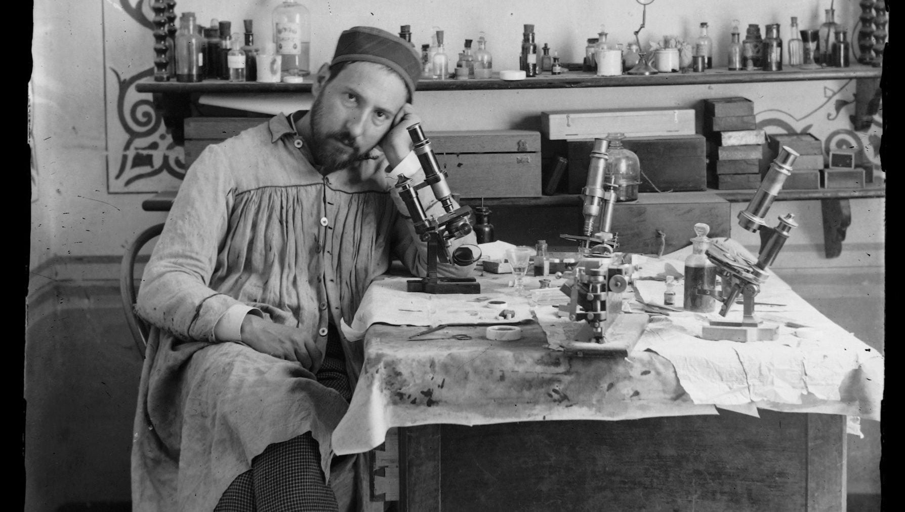
As a young man, Cajal began looking at brains of all kinds—a one-month-old human infant, a man shortly after drowning, a pigeon, a rabbit—under a microscope, and sketching what he saw. He originally wanted to be an artist, but he pursued a medical degree at the urging of his father, who was a doctor. Nevertheless, he remained fascinated by the aesthetic aspects of his scientific career. As he put it in his 1917 autobiography, Recuerdos de mi vida:
Like the entomologist hunting for brightly colored butterflies, my attention was drawn to the flower garden of the gray matter, which contained cells with delicate and elegant forms, the mysterious butterflies of the soul, the beating of whose wings may some day (who knows?) clarify the secret of mental life.
His drawings, made from roughly 1890 up until his death in 1934, so effectively illustrate now-basic neurological concepts that they are still used in neuroscience textbooks today.
Last year, 80 of his works made their US art-gallery debut in a traveling exhibit called “The Beautiful Brain: The Drawings of Santiago Ramón y Cajal” at the Wiesman Art Museum in Minneapolis, Minnesota. The drawings have traveled to the Belkin Gallery at the University of British Columbia, in Canada, and to the Grey Art Gallery at New York University, in New York City. They’ve since moved on to the MIT museum in Boston, Massachusetts, where they are on display through the end of 2018.
The appeal of these drawings go beyond the technical information they carry; they are aesthetically wonderful. The images introduce us to the astonishing reality that we carry around lush, strange landscapes inside our heads. Nearly all the drawings have analogues in non-human nature: Some resemble kelp forests, collections of coral, or roots of terrestrial trees. Others look like open flowers.
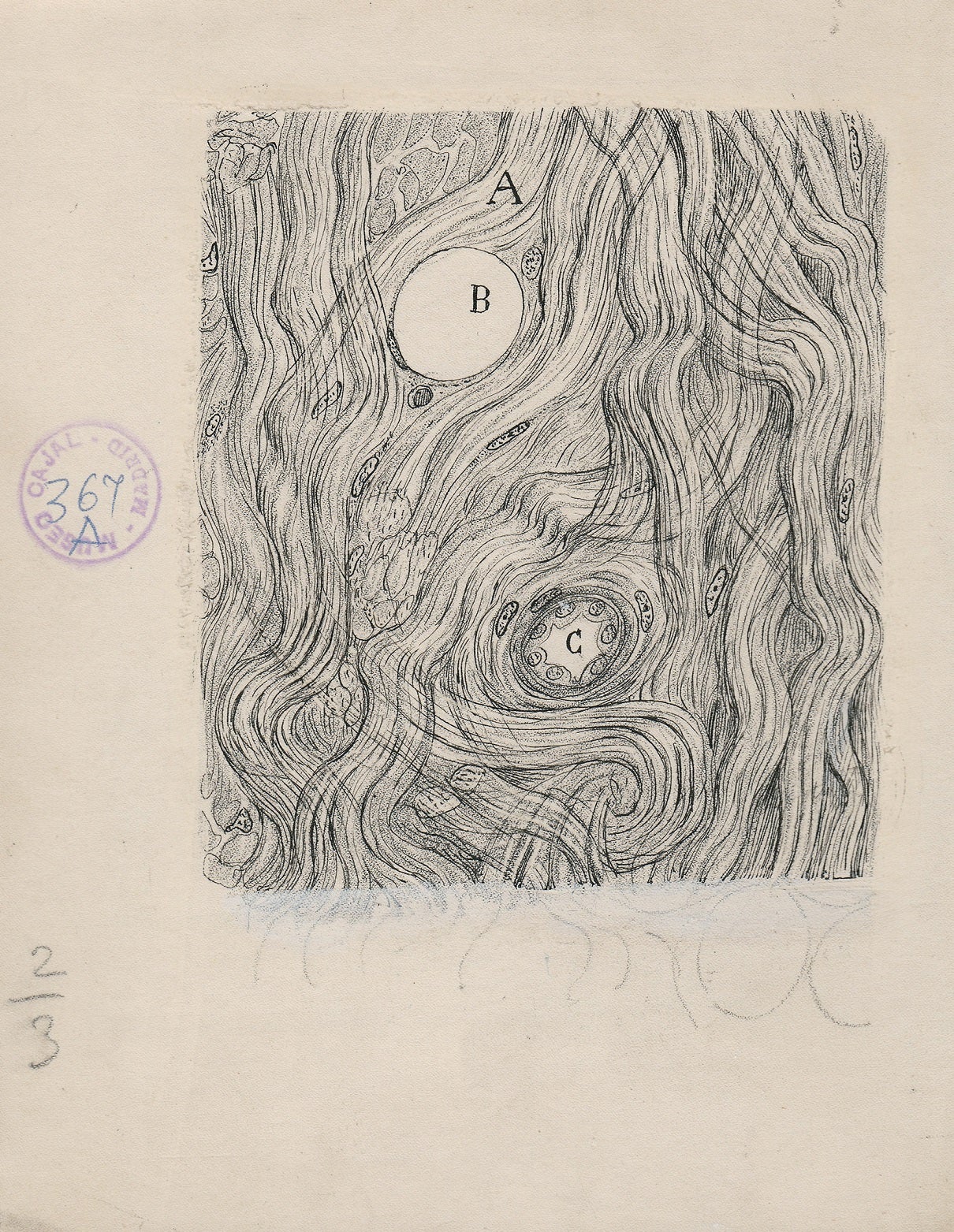
Take, for example, the root-like structure of a neuron in the cerebral cortex, the part of the brain that, among other things, receives and processes sensory information and commands motor activity:
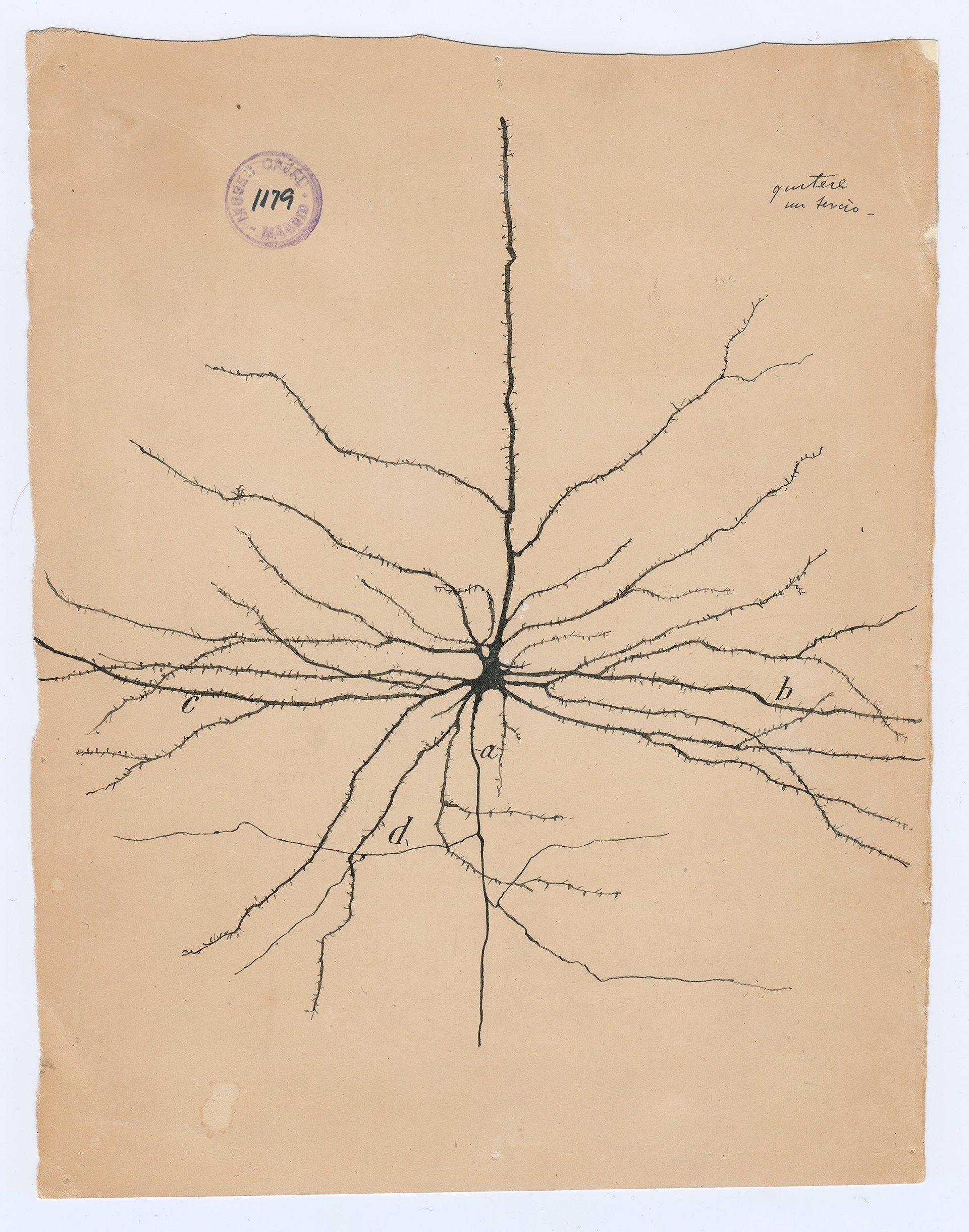
Cajal was fascinated by the brain’s botanical connections: “The cerebral cortex is similar to a garden filled with innumerable trees,” he wrote in 1894. “[T]he pyramidal cells, which can multiply their branches thanks to intelligent cultivation, send their roots deeper, and produce more exquisite flowers and fruits every day.”

“Even from the aesthetic point of view, the nervous tissue contains the most charming attractions,” Cajal wrote in his autobiography. “In our parks are there any trees more elegant and luxurious than the Purkinje cell from the cerebellum?”
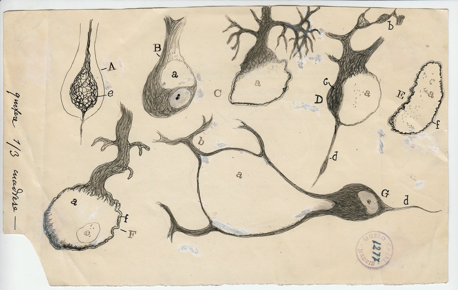

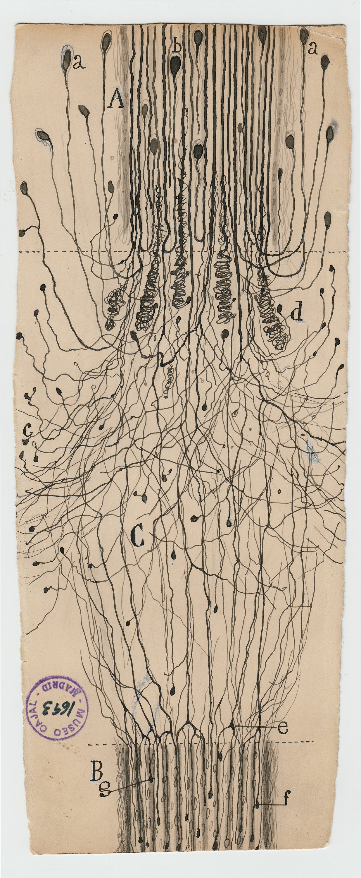
The drawing below shows a nerve partially severed from a spinal cord. From The Beautiful Brain exhibit:
At the top is the central stump (marked A), the one still attached to the spinal cord, while the disconnected end of the nerve (B) is at the bottom. The axons of the central stump grow in somewhat random directions. Some of these axons (f and g) have made it across to the disconnected end of the nerve and will regrow through the nerve all the way back to their target organ. Although neurons in the brain cannot heal after injury, this drawing shows that injured nerves outside the brain and spinal cord can.
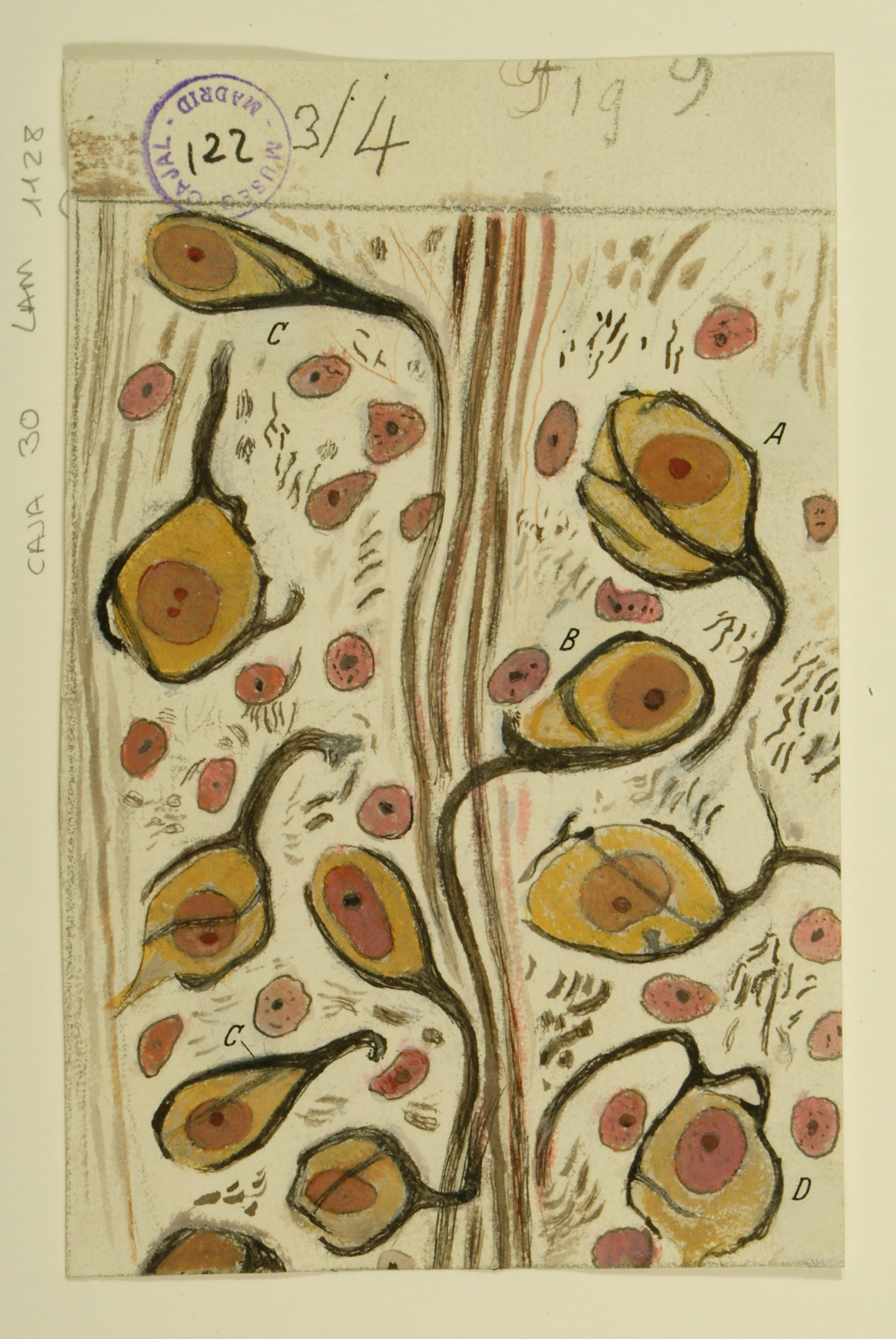
Sometimes, Cajal drew in color. Below, for example, is his rendering of the “calyces of Held,” which have a strong resemblance to calyxes, the parts of a plant that enclose a flower bud before it blooms. Here’s a description from the exhibit:
The calyces of Held—named for their resemblance to the calyxes of flowers—are synapses made by axons carrying auditory information and contacting neurons in a brain-stem structure called the trapezoid body. The calyces are the largest synapses in the brain. They are seen in these drawings as stout black lines wrapped around the yellow cells. As Cajal knew well, these cells are part of the brain system that perceives sound. The large synapses, which transmit information quickly and reliably, help us to accurately localize the source of sounds.